Kamagra gibt es auch als Kautabletten, die sich schneller auflösen als normale Pillen. Manche Patienten empfinden das als angenehmer. Wer sich informieren will, findet Hinweise unter kamagra kautabletten.
Kjp.or.kr2


Bae HK, et al. •
Korean J Pediatr 2016;59(6):262270http://dx.doi.org/10.3345/kjp.2016.59.5.262pISSN 1738-1061•eISSN 2092-7258
The effect of sildenafil on right ventricular remo
deling in a rat model of monocrotalineinduced
right ventricular failureHyun Kyung Bae, MD1, Hyeryon Lee, PhD1, Kwan Chang Kim, MD, PhD2, Young Mi Hong, MD, PhD1Departments of 1Pediatrics, 2Thoracic and Cardiovascular Surgery, Ewha Womans University School of Medicine, Seoul, Korea
Purpose: Pulmonary arterial hypertension (PAH) leads to right ventricular failure (RVF) as well as an
Corresponding author: Young Mi Hong, MD
increase in pulmonary vascular resistance. Our purpose was to study the effect of sildenafil on right
Department of Pediatrics, Ewha Womans Univer-
ventricular remodeling in a rat model of monocrotaline (MCT)-induced RVF.
sity School of Medicine, 1071 Anyangcheon-ro, Yangcheon-gu, Seoul 07985, Korea
Methods: The rats were distributed randomly into 3 groups. The control (C) group, the monocrotaline
Tel: +82-2-2650-2841
(M) group (MCT 60 mg/kg) and the sildenafil (S) group (MCT 60 mg/kg+ sildenafil 30 mg/kg/
Fax: +82-2-2653-3718
day for 28 days). Masson Trichrome staining was used for heart tissues. Western blot analysis and
immunohistochemical staining were performed.
Received: 14 March, 2016
Results: The mean right ventricular pressure (RVP) was significantly lower in the S group at weeks
Revised: 25 April, 2016
1, 2, and 4. The number of intra-acinar arteries and the medial wall thickness of the pulmonary
Accepted: 10 May, 2016
arterioles significantly lessened in the S group at week 4. The collagen content also decreased in heart tissues in the S group at week 4. Protein expression levels of B-cell lymphoma-2 (Bcl-2)-associated X, caspase-3, Bcl-2, interleukin (IL)-6, matrix metal oproteinase (MMP)-2, endothelial nitric oxide synthase (eNOS), endothelin (ET)-1 and ET receptor A (ERA) in lung tissues greatly decreased in the S group at week 4 according to immunohistochemical staining. According to Western blotting, protein expression levels of troponin I, brain natriuretic peptide, caspase-3, Bcl-2, tumor necrosis factor-α, IL-6, MMP-2, eNOS, ET-1, and ERA in heart tissues greatly diminished in the S group at week 4. Conclusion: Sildenafil al eviated right ventricular hypertrophy and mean RVP. These data suggest that sildenafil improves right ventricular function.
Key words: Sildenafil citrate, Pulmonary hypertension, Gene expression
Pulmonary arterial hypertension (PAH) affects all ages from newborns to adults. PAH
leads to dysfunctions of both pulmonary vasculature and the heart. Chronic PAH results in significant peripheral and proximal arterial remodeling and right ventricular failure (RVF)1).
The pathophysiology in PAH is vasoconstriction from endothelial dysfunction, and an
imbalance of vasoactive mediators. Remodeling of the pulmonary artery happens from an imbalance in the cell proliferation and apoptosis in the pulmonary vessel2).
Right ventricle (RV) adaptation and ventricular remodeling occurs after changes in
Copyright 2016 by The Korean Pediatric Society
pulmonary vasculature. Right ventricle hypertrophy (RVH) follows PAH because of compensatory mechanisms to the increased afterload. However, persistent overload results
This is an open-access article distributed under the
terms of the Creative Commons Attribution Non-
in RV dysfunction and failure3).
Commercial License (http://creativecommons.org/
Ventricular pressure-volume relationships, changes in wall thickness and geometry
licenses/by-nc/4.0/) which permits unrestricted non-
commercial use, distribution, and reproduction in any
are included in RV remodeling. Increased myocyte number, dimension and myocardial
medium, provided the original work is properly cited.
Korean J Pediatr 2016;59(6):262-270
extracellular matrix can also be noted4). RV remodeling is con-
(MCT) group of subcutaneous injection of MCT (60 mg/kg), S
nected with alteration in myocardial collagen content5). The
(sildenafil, 30 mg/kg) group of daily gavage feeding of sildenafil
larger amount of collagen is likely due to increased RV afterload
for 28 days after MCT injection. We sacrificed 6 rats per each
as well as the paracrine factors to provoke RVH5). The levels
group at weeks 1, 2, and 4.
of tumor necrosis factor (TNF)-α and collagen are increased in
Protocol approval was received by the Institutional Animal
RVF6). Increased gene expressions of fetal contractile proteins and
Care and Use Committees of Ewha Womans University the School
collagen were noted in the RV of rats7).
of Medicine of (approval number: 13-0226).
RV dilatation has been correlated with higher rates of apop-
tosis8). The primary crucial factor in the development of RVF is
We weighed rats and monitored for general activity throughout
Pharmacotherapy for PAH has been developing for the last
the research period. The wet weights of RV, left ventricle (LV)+
10 years to prevent the progress of the disease. Recently, there
septum (S) and lung were gauged and RV to LV+S ratio [RV/
has been much interest in pulmonary vasodilators in therapies
(LV+S)] was calculated for an index of RVH.
for PAH with heart failure. Inhaled nitric oxide (NO), endothelin antagonists and phosphodiesterase (PDE)-5 inhibitors have
3. Estimation of hemodynamics
usually been used as a vasodilator in PAH patients9).
We put the animals in the supine position with an arterial
Sildenafil is a selective agent that relaxes pulmonary vascu lar
pressure line (Physiological Pressure Transducer, MLT1199; AD
smooth muscle by inhibiting cyclic guanosine monophosphate
Instruments, Oxfordshire, UK). We inserted catheter in the ex ternal
(cGMP) specific PDE. Decrease in pulmonary vascular resistance
jugular vein to measure mean RVP. After estimating RVP, we
has been noted in sildenafil treatment in PAH10). Some research
measured pressure in the external carotid artery. Hemo dynamic
has shown that sildenafil improves endothelial function11) and
parameters were measured at weeks 1, 2, and 4.
reduces the pro-inflammatory cytokines such as TNF-α and interleukin (IL)-612) and inhibits the apoptosis progress in the PAH
4. Histopathologic analysis of pulmonary arteries
Neutral buffered formalin (10% formalin in 0.08M sodium
However, the mechanisms of the effect of sildenafil on PAH
phosphate, pH 7.4) was used for lung tissue fixation before
patients associated with congestive heart failure (CHF) are not
paraffin embedding. Three-μm-thick sections were made with
completely understood9,14). Sildenafil as a vasodilator may lead
Victoria blue staining. We captured more than 20 images of pul-
to decreased vascular pressure and tone in patients with CHF
monary arterioles per tissue section (diameter, 50–160 μm) at a
since vasoconstriction is contributed to an increase in ventricular
magnification of ×400 using a microscopic digital camera and
filling pressure and pulmonary venous pressure15). Sildenafil
analysis program (analySIS, Olympus Soft Imaging Solutions,
decreases pulmonary arteriolar resistance and pressure in CHF
Singapore). We measured the medial wall thickness between the
by increasing NO availability because the defective release of NO
internal and external elastic lamina from two sides of muscular
may be a mechanism for the constriction of pulmonary vessels16).
arteries (M1 and M2). The wall thickness was calculated as
There has been little research about the functional and struc-
follows: % wall thickness=(M1+M2)/diameter×100. The number
tural changes of RV in PAH. The effects of current PAH therapies
of intra acinar arteries was counted. A total of randomly selected
on the RV have not been thoroughly understood17).
microscopic fields per tissue section at a magnification (×200)
The object of this research is to evaluate the effects of RV re-
were analyzed.
modeling after sildenafil treatment in a rat model of monocrotaline
The heart tissues were stained with Masson's trichrome stain-
(MCT)-induced RVF.
ing. This stain was used for distinguishing collagen from mu-scle tissues. Collagen contents were quantified using Image J developed by the National Institutes of Health.
Materials and methods
5. Immunohistochemistry
We sliced formalin-fixed 4-μm section from paraffin embedded
Six-week-old male Sprague-Dawley rats with a weight of 200–
tissue blocks and then deparaffinizing with zylene and rehydrating
290 g, were used. All rats were housed in individual cages under
by serial dilutions of alcohol (70%–100%) were done. Heat anti-
standard conditions. RVF was revoked by subcutaneous injection
gen retrieval was achieved at 100°C for 10 minutes in micro wave
of 60 mg/kg MCT (Sigma Chemicals, St. Louis, MO, USA) melted
before incubation at 4°C overnight.
in 0.5 N HCl solution.
Primary antibodies were used for B-cell lymphoma-2 (Bcl-
The rats were divided into three groups: C (control) group, M
2)-associated X (Bax), caspase-3, Bcl-2, TNF-α, IL-6, matrix
Bae HK, et al. • The effect of sildenafil on right ventricular remodeling
metalloproteinase (MMP)-2, endothelial nitric oxide synthase
7. Statistical analysis
(eNOS), endothelin receptor A (ERA) from Santacruz Biotechno-
Results were expressed as the mean±standard deviation. A
logy, Santa Cruz (Santa Cruz, CA, USA) and endothelin (ET)-
Kruskal-Wallis test was used for the comparison of differences
1 from Abcam (Cambridge, UK). Slides were incubated with the
in the three groups and a Mann-Whitney test was used for
biotinylated secondary antibodies for 30 minutes at 4°C and then
between groups comparisons with Bonferroni correction. P value
with a streptavidin (Dako, Kyoto, Japan). Color development was
of <0.05 was considered statistically significant. SPSS ver. 14.0
accomplished using 3-amino-9-ethylcarbazole or DAB as a chro-
(SPSS Inc., Chicago, IL, USA) was used for all statistical analyses.
mogen. Densities were evaluated by using Image J and ex pressed
To determine whether immunohistochemical staining were
in arbitrary units.
statistically significant, we randomly selected five areas in each lung region of each group and measured mean staining densities.
6. Western blot analysis
The tissue was homogenized and centrifuged. The supernatant
was used for sodium dodecyl sulfate polyacrylamide gel electro-
phoresis. The proteins on the acrylamide gel were transferred to a polyvinylidene difluoride membrane (Millipore, Bedford,
1. Hemodynamic data
MA, USA) and the membranes were incubated at 4°C overnight
The mean RVP was greatly higher in the M group in compa-
with the appropriated primary antibodies for troponin I, brain
rison with the C group at week 1 (n=6 per each group, 17.00±0.00
natriuretic peptide (BNP), caspase-3, Bcl-2, TNF- α, IL-6, MMP-2,
mmHg vs. 8.50±0.71 mmHg, P<0.05), at week 2 (n=6 per each
eNOS, ERA from Santacruz Biotechnology and ET-1 from Abcam.
group, 25.33±1.53 mmHg vs. 11.00±1.00 mmHg, P<0.05), and at
Then, the membrane was incubated with horseradish peroxidase-
week 4 (n=6 per each group, 39.67±8.50 mmHg vs. 10.00±0.00
conjugated secondary antibodies for 1 hour at room temperature.
mmHg, P<0.05). RVP was significantly reduced in the S group
After the washing process the membrane was measured by a
in comparison with the M group at week 1 (n=6 per each group,
chemilu minescent reaction using an electrochemiluminescence-
13.00±1.00 mmHg vs. 17.00±0.00 mmHg, P<0.05), at week 2
detection kit system from GE Healthcare (Amersham Bioscience,
(n=6 per each group, 13.00±1.00 mmHg vs. 25.33±1.53 mmHg,
Piscataway, NJ, USA). The protein content was calculated with a
P<0.05) and at week 4 (n=6 per each group, 13.07±1.53 mmHg
Molecular Devices ELISA reader (Sunnyvale, CA, USA). Western
vs. 39.67±8.50 mmHg, P<0.05) (Table 1). There was no significant
blotting band intensity values were normalized by β-actin
change in aorta pressure (Data is not shown).
Table 1. The changes of right ventricular pressure at weeks 1, 2, and 4
The RV/BW ratio in the M group was greatly higher in com-
parison with the C group at week 2 (n=6 per each group, 0.81±0.08
Monocrotaline (M),
g vs. 0.58±0.02 g, P<0.05) and at week 4 (n=6 per each group,
1.76±0.14 g vs. 0.62±0.06 g, P<0.05). The RV weight in the S
group was greatly reduced in comparison with the M group at
week 4 (n=6 per each group, 1.36±0.30 g vs. 1.76±0.14 g, P<
0.05). The RV/LV+S showed significant increase in M group
Values are presented as mean±standard deviation.
in comparison with C group at week 2 (n=6 per each group,
*P<0.05: C vs. M at the corresponding time point. †P<0.05: M vs. S at the
corresponding time point.
0.41±0.06 g vs. 0.30±0.02 g, P<0.05) at week 4 (n=6 per each
Table 2. The changes of body and organ weight at weeks 1, 2, and 4 in 3 groups
Week
Values are presented as mean±standard deviation.
BW, body weight; RV, right ventricle; LV, left ventricle; S, septum; C, control; M, monocrotaline; S, sildenafil.
*P<0.05: C vs. M at the corresponding time point.
Korean J Pediatr 2016;59(6):262-270
group, 0.75±0.06 g vs. 0.32±0.03 g, P<0.05) after MCT injection.
group, 2.01±0.30 vs. 1.24±0.11, P<0.05). There was a significant
Although the RV/LV+S in the S group was lowered in comparison
reduction of the number of intra acinar arteries in the S group
with the M group, the result was not statistically significant (Table
in comparison with the M group at week 2 (n=6 per each group,
1.10±0.13 vs. 1.86±0.24, P<0.05) and at week 4 (n=6 per each group, 1.13±0.41 vs. 2.01±0.30, P<0.05) (Fig. 1C).
3. Histopathological changes in the lung tissues
Fully muscularized arteries were seen in the pulmonary ar-
4. Histopathological changes in the heart tissues
terioles (>10μM, <100μM) in the M group and S group at weeks
Since the Masson's Trichrome staining marks collagen-rich
2 and 4. Victoria blue staining identified inner and outer elastic
area in blue, the collagen-rich area was well visualized at week 2
layers of arteries (Fig. 1A).
and 4 (Fig. 2A) in each group. Collagen content (%) in the heart
The medial wall thickness (%) of pulmonary arterioles had
tissues significantly expanded in the M group in comparison
increased significantly in the M group in comparison with the
with the C group at week 2 (n=6 per each group, 19%±2.55%, vs.
C group at week 2 (n=6 per each group, 44.99±4.84 vs. 25.71±
8.5%±1.41%, P<0.05) and at week 4 (n=6 per each group, 30%±
7.85, P<0.05), at week 4 (n=6 per each group, 42.35±1.94 vs.
3.54% vs. 11%±2.12%, P<0.05) after MCT injection and signifi-
19.93±3.64, P<0.05). There was a significant decrease in the S
cantly reduced in the S group at week 4 (n=6 per each group,
group in comparison with the M group at week 2 (n=6 per each
25%±2.83 % vs. 30%±3.54%, P<0.05) (Fig. 2B).
group, 35.41±3.13 vs. 28.44±4.61, P<0.05) and at week 4 (n=6 per each group, 33.75±0.80 vs. 42.35±1.94, P<0.05) (Fig. 1B).
5. Immunohistochemical staining in lung tissues
The number of intra-acinar arteries significantly increased
The immunohistochemical staining assay by arbitrary unit
in the M group in comparison with the C group at week 1 (n=6
revealed significantly enhanced expression of Bax, Caspase-3,
per each group, 1.48±0.16 vs. 1.44±0.29, P<0.05), at week 2
Bcl-2, IL-6, MMP-2, eNOS, ET-1, ERA in the M group compared
(1.86±0.24 vs. 1.23±0.08, P<0.05) and at week 4 (n=6 per each
with the C group at week 4. In contrast, Bax, Caspase-3, Bcl-2,
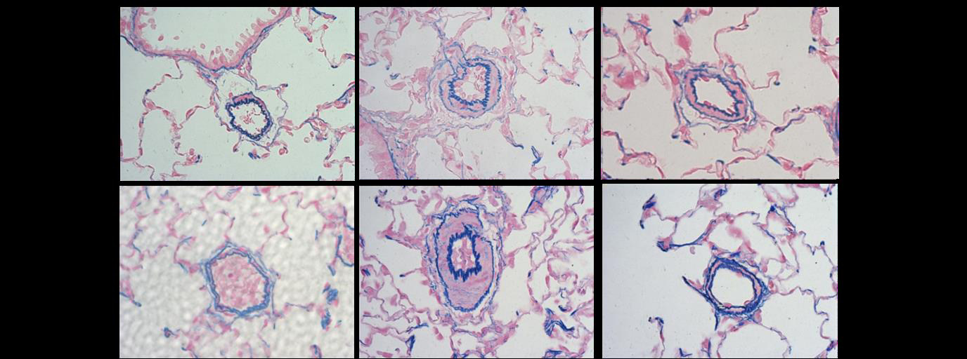
Fig. 1. Pulmonary arterioles stained with Victoria blue at the magnification of ×400 (A), medial wall
thickness (B), and the number of intra-acinar arteries (C) in the 3 groups. The medial wall thickness of
pulmonary arterioles significantly decreased in the S group in comparison with the M group at weeks
2 and 4 (panels A and B). There was a significant reduction in the number of intra-acinar arteries in
the S group in comparison with the M group at weeks 2 and 4 (panel C). C, control; M, monocrotaline;
S, sildenafil. *P<0.05: C vs. M at the corresponding time point. †P<0.05: M vs. S at the corresponding

Bae HK, et al. • The effect of sildenafil on right ventricular remodeling
Fig. 2. Masson's trichrome staining of the heart tissues (X400) (A) and collagen content (B) in the 3
groups. The collagen-rich area of heart tissue was visualized well at weeks 2 and 4 (panel A) in each
group. In comparison with group M, col agen content (%) of heart tissues significantly decreased in
the S group after 4 weeks (panel B). C, control; M, monocrotaline; S, sildenafil. *P<0.05: C vs. M at the
correspond ing time point. †P<0.05: M vs. S at the corresponding time point.
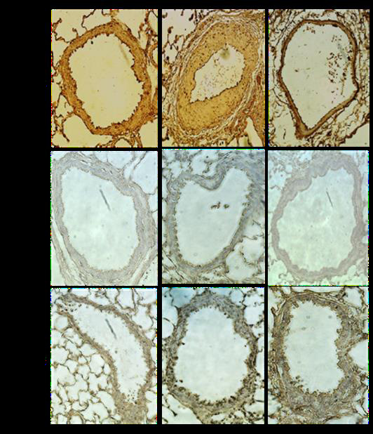
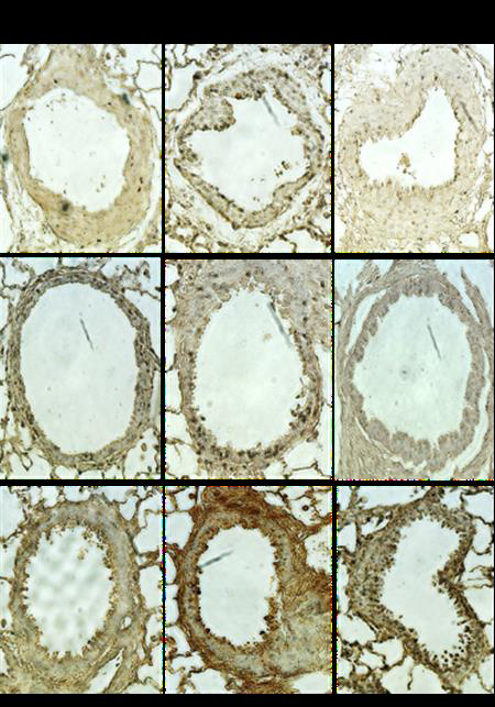
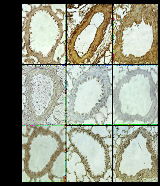
Fig. 3. Immunohistochemical staining of lung tissues (A) and relative density (B) in 3 groups at week 4.
The immunohistochemical staining assay by arbitrary unit revealed significantly reduced expressions
of Bax, Caspase-3, Bcl-2, TNF-α, IL-6, MMP-2, eNOS, ET-1, and ERA in the S group compared with the
M group at week 4 (×400). Bax, Bcl-2-associated X; Bcl, B-cell lymphoma; IL, interleukin; MMP, matrix
metalloproteinase; eNOS, endothelial nitric oxide synthase; ET, endothelin; ERA, endothelin receptor A;
C, control; M, monocrotaline; S, sildenafil. *P<0.05: C vs. M at the corresponding time point. †P<0.05:
M vs. S at the corresponding time point.
IL-6, MMP-2, eNOS, ET-1 and ERA significantly reduced in the S
MMP-2 (Fig. 4G), eNOS (Fig. 4H), ET-1 (Fig. 4I) and ERA (Fig. 4J)
group at week 4 (Fig. 3A, B).
in the M group were significantly increased in comparison with the C group at week 4. Protein expressions of troponin I (Fig. 4A),
6. Western blot analysis
BNP (Fig. 4B), Caspase-3 (Fig. 4C), Bcl-2 (Fig. 4D), TNF-α (Fig. 4E),
In western blot analyses of heart tissue, band intensity was
IL-6 (Fig. 4F), MMP-2 (Fig. 4G), eNOS (Fig. 4H), ET-1 (Fig. 4I) and
normalized to the expression of the β-actin protein at each
ERA (Fig. 4J) in heart tissues significantly lessened in the S group
week. Protein expressions of troponin I (Fig. 4A), BNP (Fig. 4B),
caspase-3 (Fig. 4C), Bcl-2 (Fig. 4D), TNF-α (Fig. 4E), IL-6 (Fig. 4F),
There was an unexpected increase in troponin I, BNP, TNF-α,
Korean J Pediatr 2016;59(6):262-270
Troponin-I
Caspase-3
Bcl-2 0.4
β-actin
β-ac 100
Fig. 4. (A-K) Western blot analysis in the heart tissue at weeks 1, 2, and 4. Protein expressions of troponin I (A), BNP (B), caspase-3 (C), Bcl-
2 (D), TNF-α (E), IL-6 (F), MMP-2 (G), eNOS (H), ET-1 (I), and ERA (J) in the heart tissues significantly lessened in the S group in comparison
with the M group at week 4. BNP, brain natriuretic peptide; Bcl, B-cell lymphoma; TNF, tumor necrosis factor; IL, interleukin; MMP, matrix
metalloproteinase; eNOS, endothelial nitric oxide synthase; ET, endothelin; ERA , endothelin receptor A; C, control; M, monocrotaline; S,
sildenafil. *P<0.05: C vs. M at the corresponding time point. †P<0.05: M vs. S at the corresponding time point.
Bae HK, et al. • The effect of sildenafil on right ventricular remodeling
and IL-6 protein expression levels in the C group. The small
sildenafil administrations in MCT rats. The protein expressions
number of animals causes the unexpected increase in protein
related to endothelial cell dysfunction such as eNOS, ET-1, and
expression levels because of individual differences.
ERA were significantly reduced after sildenafil treatment. ET and
RV dysfunction (BNP), apoptosis (caspase-3, Bcl-2), inflamma-
ERA have been noted to affect tissue remodeling. MMP-2 is an
tion (TNF-α, IL-6) and endothelial dysfunction (eNOS, ET-1,
enzyme that degrades extracellular matrix. ET-1 plays a role as a
ERA) associated genes were significantly reduced after sildenafil
vasoconstrictor and enhances cell proliferation and fibrogenesis
treatment at week 4.
by controlling MMP19). In our current research, the MMP-2 ex-pression was augmented in the M group and lessened in the S group.
NO, produced by endothelial NO synthase, acts as a vasodilator
in pulmonary vasculature. In some reports, the overproduction
In this research, RVF and PAH were developed by intraperi-
of eNOS in mice models prevents hypoxia-induced PAH20) and
toneal injection of MCT, which is selectively toxic to the endothe-
severe PAH was seen in eNOS deficient mice exposed to mild
lium of the pulmonary artery and causes PAH from 1 week after
hypoxia21). On the other hand, there is a report that sildenafil ele-
injection. We were able to confirm this by increased RVP and
vates the cGMP in target cells and then decreases NOS activity
RV/LV+septum. RVF was confirmed by signs such as tachypnea,
and plasma NO level22). In our results, eNOS protein expressions
significant edema and ascites.
were significantly increased in the M group compared with the C
RVH is the underpinning of the functional and structural
group and decreased after sildenafil treatment at week 4.
changes in RV remodeling. RV muscle mass is more susceptible
Inflammation and apoptosis are important mechanisms in the
to changes in afterload5). The functional and structural changes of
molecular change of the RVF in PAH. Increased inflammatory
cardiomyocytes happen because of stress on the myocardiocytes.
cytokines are known to increase pulmonary circulation and dilate
Myocardiocytes under stress have a long contraction time and
RV of PAH patients3). In this study, the expressions of IL-6 and
increased wall stress, following ventricular remodeling, con-
TNF-α increased in MCT induced RVF rats and decreased after
tractile dysfunction and ventricular enlargement with pressure
sildenafil treatment. In addition, the failing RV may be subject to
overload. Myocardiocyte changes include lengthened RV myo-
cardiomyocyte apoptosis13). There was also the result that the ex-
cyte diameter, expanded mean myocardial cell volume, elongated
pressions of apoptosis related proteins, including caspase-3, bcl-2
myocyte length with shrunk cross-sectional areas and extended
augmented in the M group and reduced in the S group. Induction
extracellular space5).
of apoptosis is another example of the cellular changes associated
However, the critical mechanisms of change from hypertrophy
with RV remodeling. Bussani et al.8) compared apop tosis in myo-
to dilatation to cause RVF in PAH have not been well investi-
cardium with the degree of unfavorable cardiac remodeling.
gated3,18). Although incremented afterload is the first causal factor
Apop tosis may be the primary critical factor to cause biventricular
for RV adaptation in PAH17) neurohormonal signaling, oxidative
stress, inflammation, ischemia, apoptosis and endothelial
In our research, protein expressions of troponin I and BNP in
dysfunction may induce the development of RV dilatation and
the heart tissues significantly decreased at week 4 after sildenafil
treatment. The troponin is one of the myocardial regulatory
In our current research, increased inflammation (TNF-α and
proteins and may be related to the pathobiology of heart failure
IL-6 expressions), apoptosis abnormality (caspase-3 and Bcl-
23,24). Contractile dysfunction in heart failure is related to changes
2 expressions), endothelial dysfunction (eNOS, ET-1, and ERA
in regulatory and contractile and protein expression.
expressions) and cardiac dysfunction (BNP and Troponin I
Ventricular BNP level is upregulated in cardiac failure and
expressions) were found in our MCT induced RVF model.
locally in the area around a myocardial infarct. BNP is valuable
Collagen change has also been reported as an important patho-
in the diagnosis and prognosis of heart failure. It is regarded
physiology in RV remodeling. Heart failure related to myocardiac
to be the best biomarker in heart failure25). BNP is released by
infarction in a rat model showed an increased level of collagen
myocardial stretch from RV overload.
gene expressions in the RV7). The increased levels of RV collagen
Hemodynamic effect of PDE-5 inhibitor on RVF with PAH has
and TNF-α were seen in failing hearts of patients with end-stage
been recently found26). PDE-5 inhibitors may contribute to sup-
car diomyopathy6).
press RVH and improve RV function as well as decrease the RV
In our study, collagen content in the heart tissues significantly
afterload27). PDE-5 has been reported to be prominently upre-
increased at weeks 2 and 4 in MCT rats and significantly de-
gulated in RVH myocardium4).
creased after sildenafil treatment.
Sildenafil as a selective PDE-5 inhibitor is also known to pre-
We investigated changes of RV protein expression levels after
vent RV remodeling by a complex mechanism like modulating
Korean J Pediatr 2016;59(6):262-270
cGMP and calcium signaling28). Sildenafil increases cardiac
tute (2014) and Basic Science Research Program through the
index26) and prevents progressive chamber dilation, dysfunction
National Research Foundation of Korea (NRF) funded by the
and fibrosis of the heart with pre-existing hypertrophy and forces
Ministry of Education (2013R1A1A3004619).
the contractility of cardiomyocytes28).
Sildenafil has also been reported to decrease pulmonary
vascular resistance (PVR) and mean pulmonary artery pressure
in patients with heart fail ure and PAH29). In our study, there was a significant decrease of RVP after sildenafil treatment. There
1. Chesler NC, Roldan A, Vanderpool RR, Naeije R. How to measure
were also improvements in RVH by estimating the decrease of
pulmonary vascular and right ventricular function. Conf Proc IEEE Eng Med Biol Soc 2009;2009:177-80.
RV weight in addition to improvement in pathology and gene
2. Wilkins MR. Pulmonary hypertension: the science behind the
expressions of the lung and heart.
disease spectrum. Eur Respir Rev 2012;21:19-26.
Sildenafil may include the various and valuable combination
3. Vonk-Noordegraaf A, Haddad F, Chin KM, Forfia PR, Kawut SM,
of inotropic, antihypertrophic27) and afterload-decreasing effects
Lumens J, et al. Right heart adaptation to pulmonary arterial hypertension: physiology and pathobiology. J Am Coll Cardiol
on the RV without significantly influencing systemic hemody-
4. Nagendran J, Archer SL, Soliman D, Gurtu V, Moudgil R, Haromy
We considered 30 mg/kg of sildenafil was the optimal dose
A, et al. Phosphodiesterase type 5 is highly expressed in the hyper-
since the oral administration of sildenafil dose from 30 mg/kg to
trophied human right ventricle, and acute inhibition of phospho-
100 mg/kg in rats seemed to be the effective regarding clearance.
diesterase type 5 improves contractility. Circulation 2007;116:238-48.
Meanwhile, we assessed that the dose of sildenafil below 30
5. Kret M, Arora R. Pathophysiological basis of right ventricular
mg/kg is less effective than the dose of silde nafil of 30 mg/kg
remodeling. J Cardiovasc Pharmacol Ther 2007;12:5-14.
considering the short half life according to previous data31).
6. Kucuker SA, Stetson SJ, Becker KA, Akgul A, Loebe M, Lafuente
Our gene expression studies of immunohistochemistry provided
JA, et al. Evidence of improved right ventricular structure after LVAD support in patients with end-stage cardiomyopathy. J Heart
that the expressions of Bax, caspase-3, Bcl-2, IL-6, MMP-2,
Lung Transplant 2004;23:28-35.
eNOS, ET-1, and ERA significantly decreased in the S group at
7. Yoshiyama M, Takeuchi K, Hanatani A, Shimada T, Takemoto Y,
week 4. In this study, we demonstrated that sildenafil improves
Shimizu N, et al. Effect of cilazapril on ventricular remodeling
endothelial dysfunction, apoptosis, inflammation and remodeling
assessed by Doppler-echocardiographic assessment and cardiac gene expression. Cardiovasc Drugs Ther 1998;12:57-70.
in the lung and heart tissues of MCT-induced rats.
8. Bussani R, Abbate A, Biondi-Zoccai GG, Dobrina A, Leone AM,
However, further research is required to identify the exact me-
Camilot D, et al. Right ventricular dilatation after left ventricular
chanism of sildenafil effect. This study has some limitations. A
acute myocardial infarction is predictive of extremely high peri-
longitudinal follow-up study is needed. Our study ended at week
infarctual apoptosis at postmortem examination in humans. J Clin Pathol 2003;56:672-6.
4 because the survival rate in the M group was 56%, which is
9. Lepore JJ, Maroo A, Bigatello LM, Dec GW, Zapol WM, Bloch
lower than that of the C group (100%) and the S group (100%) at
KD, et al. Hemodynamic effects of sildenafil in patients with con-
week 4. There is also a lack of comprehensive assessment as to
gestive heart failure and pulmonary hypertension: combined
the diverse molecular changes involved in the effect of sildenafil
administration with inhaled nitric oxide. Chest 2005;127:1647-53.
10. Galie N, Ghofrani HA, Torbicki A, Barst RJ, Rubin LJ, Badesch D, et
al. Sildenafil citrate therapy for pulmonary arterial hypertension.
In conclusion, sildenafil treatment improved mean RVP, RVH
N Engl J Med 2005;353:2148-57.
and ventricular remodeling after sildenafil treatment. These
11. Balarini CM, Leal MA, Gomes IB, Pereira TM, Gava AL, Mey relles
results may have important implications for the experimental and
SS, et al. Sildenafil restores endothelial function in the apolipo-protein E knockout mouse. J Transl Med 2013;11:3.
therapeutic use of sildenafil.
12. Toward TJ, Smith N, Broadley KJ. Effect of phosphodiesterase-5
inhibitor, sildenafil (Viagra), in animal models of airways disease. Am J Respir Crit Care Med 2004;169:227-34.
Conflict of interest
13. Schwartz BG, Levine LA, Comstock G, Stecher VJ, Kloner RA.
Cardiac uses of phosphodiesterase-5 inhibitors. J Am Coll Cardiol 2012;59:9-15.
No potential conflict of interest relevant to this article was
14. Urbanowicz T, Straburzyńska-Migaj E, Katyńska I, Araszkiewicz
A, Oko-Sarnowska Z, Grajek S, et al. Sustained improvement of clinical status and pulmonary hypertension in patients with severe heart failure treated with sildenafil. Ann Transplant 2014;19:325-30.
15. Guazzi M, Tumminello G, Di Marco F, Guazzi MD. Influences of
sildenafil on lung function and hemodynamics in patients with
This research received support from the Korean Medical Insti-
chronic heart failure. Clin Pharmacol Ther 2004;76:371-8.
Bae HK, et al. • The effect of sildenafil on right ventricular remodeling
16. Moraes DL, Colucci WS, Givertz MM. Secondary pulmonary
dium. Heart Fail Rev 2005;10:199-209.
hypertension in chronic heart failure: the role of the endothelium
25. Panagopoulou V, Deftereos S, Kossyvakis C, Raisakis K, Gianno-
in pathophysiology and management. Circulation 2000;102:1718-
poulos G, Bouras G, et al. NTproBNP: an important biomarker in
cardiac diseases. Curr Top Med Chem 2013;13:82-94.
17. Bogaard HJ, Abe K, Vonk Noordegraaf A, Voelkel NF. The right
26. Michelakis E, Tymchak W, Lien D, Webster L, Hashimoto K, Archer
ventricle under pressure: cellular and molecular mechanisms of
S. Oral sildenafil is an effective and specific pulmonary vasodilator
right-heart failure in pulmonary hypertension. Chest 2009;135:94
in patients with pulmonary arterial hypertension: comparison with
inhaled nitric oxide. Circulation 2002;105:2398-403.
18. McMurtry MS, Archer SL, Altieri DC, Bonnet S, Haromy A, Harry
27. Takimoto E, Champion HC, Li M, Belardi D, Ren S, Rodriguez ER,
G, et al. Gene therapy targeting survivin selectively induces pul-
et al. Chronic inhibition of cyclic GMP phosphodiesterase 5A pre-
monary vascular apoptosis and reverses pulmonary arterial hyper-
vents and reverses cardiac hypertrophy. Nat Med 2005;11:214-22.
tension. J Clin Invest 2005;115:1479-91.
28. Nagayama T, Hsu S, Zhang M, Koitabashi N, Bedja D, Gabrielson
19. Abraham D, Ponticos M, Nagase H. Connective tissue remodeling:
KL, et al. Sildenafil stops progressive chamber, cellular, and mole-
cross-talk between endothelins and matrix metalloproteinases.
cular remodeling and improves calcium handling and function in
Curr Vasc Pharmacol 2005;3:369-79.
hearts with pre-existing advanced hypertrophy caused by pressure
20. Ozaki M, Kawashima S, Yamashita T, Ohashi Y, Rikitake Y, Inoue N,
overload. J Am Coll Cardiol 2009;53:207-15.
et al. Reduced hypoxic pulmonary vascular remodeling by nitric
29. Lewis GD, Shah R, Shahzad K, Camuso JM, Pappagianopoulos PP,
oxide from the endothelium. Hypertension 2001;37:322-7.
Hung J, et al. Sildenafil improves exercise capacity and quality of
21. Fagan KA, Fouty BW, Tyler RC, Morris KG Jr, Hepler LK, Sato K,
life in patients with systolic heart failure and secondary pulmonary
et al. The pulmonary circulation of homozygous or heterozygous
hypertension. Circulation 2007;116:1555-62.
eNOS-null mice is hyperresponsive to mild hypoxia. J Clin Invest
30. Webster LJ, Michelakis ED, Davis T, Archer SL. Use of sildenafil for
safe improvement of erectile function and quality of life in men
22. Sirmagul B, Ilgin S, Atli O, Usanmaz SE, Demirel-Yilmaz E.
with New York Heart Association classes II and III congestive heart
Assessment of the endothelial functions in monocrotaline-induced
failure: a prospective, placebo-controlled, double-blind crossover
pulmonary hypertension. Clin Exp Hypertens 2013;35:220-7.
trial. Arch Intern Med 2004;164:514-20.
23. Adamcova M, Sterba M, Simůnek T, Potacova A, Popelova O, Gersl
31. Shin HS, Bae SK, Lee MG. Pharmacokinetics of sildenafil after
V. Myocardial regulatory proteins and heart failure. Eur J Heart
intravenous and oral administration in rats: hepatic and intestinal
Fail 2006;8:333-42.
first-pass effects. Int J Pharm 2006;320:64-70.
24. VanBuren P, Okada Y. Thin filament remodeling in failing myocar-
Source: http://kjp.or.kr/upload/KJP_59_6_262_270_20125550548.pdf
Your Health Reimbursement Arrangement (HRA) dollars may be able to be used to pay for co-payments, co-insurance, and deductibles. But that's not all. You may also be able to use your HRA money to pay for many expenses in the following categories: Medical, Dental Care, Eye Care, and Over-the-Counter (OTC) medications. Eligible items can vary by employer, so check the specifics of your particular HRA plan.
Study of Bacteriological Profile and Antibiotic Sensitivity & Resistance Pattern in Pus Culture Isolates at Tertiary Care Teaching Hospital in Bhopal Nidhi Jain, Abhay Joshi, Rakesh Sonawane, Arun Srivastav Objective: To study the bacteriological profile and the pattern of antibiotic sensitivity and resistance in pus culture isolates in a tertiary care teaching hospital in Bhopal. Materials and Methods: Pus specimens submitted to the microbiology laboratory for routine cultures and sensitivity were analyzed prospectively for the duration of 2 months. Antimicrobial susceptibility testing was performed by Kirby-Bauer Disk Diffusion method. Results: Among the total 440 samples analyzed, 319 samples (72.5%) were found to be positive for growth. Out of 319 growths, 286 shows single isolate and 33 shows mixed (double) isolates. Out of these positive samples, 269 samples (84.33%) were positive for