Kamagra gibt es auch als Kautabletten, die sich schneller auflösen als normale Pillen. Manche Patienten empfinden das als angenehmer. Wer sich informieren will, findet Hinweise unter kamagra kautabletten.
Ninth arab conference on the peaceful uses of atomic energy, beirut, 3-6 december 2008
Original article
Epidemiological Studies on Strongyloides stercoralis at Dilla
District, Ethiopia
Feleke Eriso
Parasitologist, Department of Biology, Dilla University, Dilla, Ethiopia
Corresponding Author
Background and study aim: Some
Results: In the study a total of 710 student
Feleke Eriso
children were examined for
Strongyloides
parthenogenesis or asexual reproduction
stercoralis infection out of whom 142
and hermaphroditism (protandrogony) to
(in 1st study) were positive, confirming
+251916514682
be the only mode of reproduction of
the infection rate to be 20% or 198
parasitic female
Strongyloides stercoralis
positive (in 2nd study) the infection rate
in human hosts as parasitic males of it did
being 28% by the parasite. Then, the
not exist in human hosts. Therefore, the
average infection rate was
first objective was to work out the
developmental stages and sexes of the
infection rate of
Strongyloides stercoralis
parasite were obtained in the study
in the population of elementary schools
children at Dilla district; secondly,
Conclusion: The presence of many
to produce a visible evidence for the prese
parasitic males with everted spicules
nce of many parasitic males of
Strongyloi-
observed in fresh stools samples during
stercoralis; Diagnosis; Morbidity;
des stercoralis as there are parasitic femal
this study had been a very strong evidence
Mortality; Treatment;
-es in fresh stools samples of human
for the fact that male and female
Immunocompetent
hosts; and thirdly, to replace the unfit
copulation & fertilization were naturally
term by a correct one.
taking place among parasitic worms of
Strongyloides
stercoralis
Patients and methods: Stools samples
hosts. Parasitic and free living males of
S.
were collected from student children of
stercoralis have the same morphology
elementary schools, and observed under
including the curved or coiled posterior
parasitology after employing Baermann
apparatus technique.
article on the internet which stated
that the parasitic generation of
S.
Strongyloides stercoralis is known to
stercoralis was known not to have
have two life cycles: an internal sexual
parasitic males and the parasitic
cycle, involving parasitic worms that
females used to reproduce only by the
constitute the parasitic generation, and
asexual method of reproduction [8]. In
the parasitic generation, when the
interacting among free-living worms
filariform larvae are in contact with
that represent the free-living generation
cutaneous blood vessels and are
stated by authors in modern textbooks,
carried through the right heart to the
journals, and on the internet that the
lungs [9]. Then, sexually mature
type of reproduction in the parasitic
parasitic females settle in the tissues
generation of
S. stercoralis in human
of epithelial mucosa to lay eggs that
hosts was only by parthenogenesis of
hatch soon and are discharged in the
parasitic females in the absence of
stools each day [10,11]. When all or
parasitic males [4-7]. Due to this
metamorphose into
parasitic male had been omitted in the
figures that demonstrated the life
be onset by invading the
cycle of the parasitic generation of
S.
of the ileum or colon, travel to lungs a
stercoralis in all modern and relevant
nd then return to the intestine to
textbooks, journals, and on the
mature in the mucosa [12-15].
internet. Not only that, there was an
Eriso, Afro-Egypt J Infect Endem Dis 2014; 4(2): 69-79
Original article
Disseminated strongyloidiasis had been reported
Strongyloidiasis [19-21]. Some of the clinical
in both of two recipients of kidney allografts
presentations of strongyloidiasis can be high-
from a single cadaver donor [16]. It was
also reported that in a 53-year-old man who had
Cutaneous with larva currens (racing larvae),
lung cancer, fulminantly fatal strongyloidiasis
pruritic linear or serpiginous, creeping urticarial
had developed following postchemotherapy of
eruption, dermatologic lesions, and petechiae;
immunosuppression, resulting in the death of the
Pulmonary with persistent wheezing, cough,
patient within 48 hrs [16]. The development of a
and deteriorating respiratory status; and
florid strongyloidiasis was observed in a 45-year-old man, following anticancer chemotherapy
Intestinal with vomiting, abdominal pain,
when eggs of
S. stercoralis were seen in the
watery diarrhea and constipation.
stools [17]. One scientific study has reported that
almost all deaths due to helminths in the United States result from
S. stercoralis hyperinfection
PATIENTS AND METHODS
mortality rates because the occurrence of
The suitable type of study selected to answer the
hyperinfection can be as high as 87% [18].
question of this research work was the Cross Sectional Study. The statistical methods pre-
Aim of the study
planned to be employed in analyzing and
The aim of the study has three objectives to work
interpreting the results were the expression by
percentage and standard deviation.
First, to determine the infection rate of
S.
stercoralis in the population of elementary
Eight different elementary schools found at Dilla
schools children at Dilla district;
district were selected to be the sites of fresh stools sample collection from student children. It
Second, to produce a visible evidence for the
presence of many parasitic males of
S.
geographical points of view, to notify that Dilla
stercoralis as there are parasitic females in
district is located in Gedio Zone that is found in:
fresh stools samples of human hosts; and
Southern Ethiopia,
Thirdly, to replace the unfit morphologic term
The continent of Africa, and
by a correct one. Is parthenogenesis or asexual
The northern hemisphere between the tropic of
reproduction true in the parasitic generation of
cancer and the equator.
S. stercoralis in human hosts?
Concerning some morphological features of this
documenting related information from the
parasite, the part of the worm's body that is
schools had been carried out from 6/12/2006 to
known as the tail is the posterior part of body
10/06/2007 and again repeated in depth from
beginning from cloaca in the parasitic males or
10/9/2008 to 25/6/2009. However, the deliberate
follow up to be certain about the prevalence and
females. Cloaca is the opening through which
persistence of the parasite at Dilla district, was
spicules are everted at times of copulation &
performed by taking fresh stools samples of ten
fertilization and it is also the outlet of
students from each of the eight different
the digestive tract. The 3 stages of human
elementary schools every year from 2000 up to
strongyloidiasis are Intestinal Strongyloidiasis,
the beginning of 2014.
Gastropulmonary Strongyloidiasis and Disseminated
Eriso, Afro-Egypt J Infect Endem Dis 2014; 4(2): 69-79
Original article
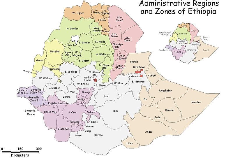
Figure (1): Map of administrative regions and zones of Ethiopia. Gedio
Specific sample size:
including the participation of the child for the
The sample size taken from the participant
necessary information, with the exception of the
student children was 710. Each day, Monday
column under S. stercoralis, because it had to be
through Friday (i.e. every week), fresh stools
filled either "–" or "+" for S. stercoralis, by the
samples, of ten student children were taken to
researcher after examining the fresh stools
parasitology laboratory of Dilla University.
sample. Writing the name of the student child in
When the sample was taken from the student
the steps of raw data collection was important to
child, he/she gave fresh stools sample in a bottle
identify the child for giving treatment if he/she
on which his/her I.D. No. was written. In
had been found to be positive for the parasite,
addition to this, on that very day and moment a
because many children could not remember their
table that had columns with the headings of Date,
I.D. No. Of course, it was decided not to write
Name of Child, I.D. No. of child, Class (grade),
the name of the student child in the report of the
Age in Year, Sex, S. stercoralis, Education of
parents, and Job of parents were filled by
Eriso, Afro-Egypt J Infect Endem Dis 2014; 4(2): 69-79
Original article
Diagnostic Examination:
No. of the student child from whom the fresh
The diagnostic examination of fresh stools
stools sample was taken to be examined.
sample of each student child involved the
These seven steps were repeated for the fresh
following nine steps.
stools sample of each of the remaining 9
Baermann funnel apparatus was constructed
student children.
and the lower opening of the rubber tubing
Using a dropper, a drop of suspension was
fitted to the stem of the funnel was closed.
taken from the surface of bottom sediment of
Water warmed to 400 C was poured into the
the labeled test tube suspension and placed on
funnel of the Baermann apparatus and the
a clean glass slide and then covered with a
cheese cloth, that contained the fresh stools
cover slip. The preparation was examined
sample of the student child and tied with its
under the low power objective of a research
peripheral edges to the rim of the funnel, was
microscope to confirm the presence or
partially immersed in the water warmed to 400
absence of S. stercoralis in the fresh stools
C. This was done because if adults as well as
sample of the student child. The sample of
juveniles of S. stercoralis were present in the
each student child was examined in this way.
stools, they would be attracted by the warm
The column under the heading of S.
temperature of water (about 37.5o C as there
stercoralis for each student child was marked
was dissipation of heat from the initial 40o C
"–" indicating the absence or "+" confirming
of the added water to the surrounding
the presence of S. stercoralis in the fresh
materials and equipment) and escape into the
stools sample taken.
warm water through the pores of the cheese
The suspensions positive for S. stercoralis
were fixed and preserved by adding 10%
After staying 1 hour and 30 minutes, the
formaldehyde. Each container bottle of
closed lower end of the rubber tubing was
preservation in 10% formaldehyde was labeled S.
opened, releasing the water found in the funnel of
stercoralis larvae/other stages including the
Baermann apparatus into a 500 ml beaker. The
date of collection and kept in a safe place in
stools left behind in the cheese cloth was
the laboratory of parasitology. Water emergence
thrown into the tube of toilet after being
semi-concentration technique for detecting
treated with a disinfectant (iodine solution) and
strongyloides larvae in feces was also used
washed away by a current of water.
when there were needs to supplement the
The water released and collected in the 500 ml
Baermann method [22].
beaker was centrifuged at a speed of 1000 rpm (revolutions of the rotor per minute) for 2
Water emergence semi-concentration technique
minutes using a manual centrifuge loaded with
for detecting S. stercoralis larvae in feces:
4 centrifuge tubes and anchored to the edge of
A fresh (not more than 2 hours old) formed or
semi-formed fecal specimen is required. The
From each centrifuge tube the supernatant was
method is as follows:
poured off into a waste collecting bucket to be
Using a piece of stick, make a central
thrown into the tube of toilet drainage line by
depression in the specimen contained in a
treating with the disinfectant.
vial or bottle. Fill the depression with warm
Using a dropper, about 2 ml of the supernatant
water (about 37.50ºC).
was added to the sediment of one of the 4
Incubate the specimen in a 35-37.50ºC
sediment containing centrifuge tubes and
incubator for 1.5 to 3 hours during which time
shaked well by closing its mouth with its own
the larvae will migrate out of the feces into the
fittingly tight lid. The action of shaking was to
change the sediment into a transferable
Using a plastic bulb pipette or Pasteur pipette,
suspension. The same suspension was
transfer some of the water to a slide and cover
Transferred to each of the remaining 3
with a cover glass. Alternatively, transfer all
centrifuge tubes one by one where in each
the water to a conical tube, centrifuge, and
case the centrifuge tube was shaked well and
transfer the sediment to a slide.
the sediment was changed into suspension.
Examine the preparation, under the low or
Next, the sediment collected in the form of
middle power objective lens of a compound
suspension from 4 centrifuge tubes was
light microscope, for motile larvae of S.
poured into a test tube labeled with the I.D.
stercoralis.
Eriso, Afro-Egypt J Infect Endem Dis 2014; 4(2): 69-79
Original article
Treatment :
child, infected with S. stercoralis, before he
The drug, that was available to treat the student
had been given treatment.
children infected with S. stercoralis and ordered
Excess water was added to the topsoil of all
by the medical doctor assigned to assist the
the three petridishes and were incubated at 280
researcher of this study, was albendazole (Avion:
C on the same day.
Nabros, England) in the study project of
The topsoil of petridish No. 1, 2, and 3 were
6/12/2006 to 10/06/2007. On the other hand, the
examined, using Baermann funnel apparatus
drug of choice ordered by the medical doctor in
technique to check the growth of free-living
the study of 10/09/2008 to 25/06/2009 was
generation of S. stercoralis, after 11, 30, and
ivermectin (Ochoa: Ravenbhel, India).
48 days of initial incubation respectively.
The worms of the free-living generation of S.
The dose of albendazole:
stercoralis collected from the three petridishes
Each infected child whose age was 9 years and
of topsoil using Baermann techniquewere
above was advised to take two albendazole
fixed and preserved in 10% formaldehyde to
tablets at one time after dinner immediately
be used for the preparation of permanent
before going to bed for night sleep daily for two
slides [5].
consecutive days whereas those whose ages were 8 years and below were given 1 bottle (20 ml)
Method of Safranin stain preparation :
albendazole oral suspension to take after dinner
I.1. Safranin O stock solution: Dissolve 2.5g
immediately before going to bed for night sleep
safranin O Certistain in 100 ml of 96%
daily for three consecutive days. It was notified
ethanol. This is a stock solution.
that each tablet contained 200 mg albendazole
2. For use: 10 ml of stock solution should be di
USP whereas each bottle (20 ml) contained 400
luted with 90 ml of distilled water [23,24].
mg albendazole USP.
OR II. 1. Safranin powder……………0.1 g
The dose of ivermectin:
2. Distilled water…………….100 ml
The prescription was stated as follows in
The safranin powder is dissolved in the
proportion to individual student child's body
distilled water measured above.
weight. (Note: in this particular ivermectin 1 tablet is 6 mg in weight).
Preparation
permanent
microphotographs :
6 mg tablet of ivermectin
In short, the preparation of S. stercoralis
15-24 kg 0.5 tablet, single dose on empty stomach.
permanent slides was effectively done by
25-35 kg 1 tablet, single dose on empty stomach
applying the following Yetwin mounting
36-50 kg 1.5 tablets, single dose on empty stomach
medium [25].
51-65 kg 2 tablets, single dose on empty stomach
2.5 tablets, single dose on empty stomach
Yetwin Mounting Medium:
i 1. 10% bacto-gelatin, granular, aqueous
Each student child was advised to take the
2. Glycerin 50.0 ml
tablet/s with a glass of water in the morning after
3. 1% chromium potassium sulfate 100.0 ml
waking up from bed and begin taking meal at
aqueous (Chromium)
4. Phenol (carbolic acid), melted
Growth of free-living generation of S. stercoralis
ii. Gelatin was dissolved in boiling water (i.e., a
in the autoclaved topsoil in petridish incubated at
400 ml beaker, into which 10 g of gelatin &
90 ml of pure water were added, was
Topsoil that contained organic substance was
immersed in a volume of boiling water in a
taken and put into three different petridishes.
larger heat-resistant dish) and glycerin was
Each of the petridishes was closed with its own
added to it. After mixing glycerin and 10%
lid and labeled 1, 2, and 3.
gelatin solution, 1% chromium potassium
sulfate solution and phenol were added to the
Next, the petridishes with their contents of
mixture of glycerin and 10% gelatin solution.
topsoil were autoclaved.
The medium was liquefied in 15 minutes at
The topsoil autoclaved in each of the Petri-
dishes was inoculated with S. stercoralis from fresh stools sample obtained from a student
Eriso, Afro-Egypt J Infect Endem Dis 2014; 4(2): 69-79
Original article
iii. Thereafter, the S. stercoralis worms were
alcoholism; asthma; tuberculosis; malnutrition;
transferred from 10% formaldehyde directly
chronic pulmonary disease; leprosy; chronic renal
into a drop of mounting medium, placed on a
failure; impaired bowel motility; immuno-
clean slide. The mounting medium with the
suppressive therapy for diseases such as
worms was covered with a cover slip.
rheumatic disease, malignancy or cancer, and organ transplants; and promiscuous defecation.
iv. Then, within overnight the gelatin hardened
to form a permanent slide of S. stercoralis
The difference in the infection rate of S.
stercoralis in children due to the difference in the status of environmental sanitation & economic
From the permanent slides prepared microphoto-
income in the families' residence areas of the
graphs of the larvae and other stages of S.
children was analyzed by the statistic of standard
stercoralis were taken using a digital camera
deviation. In this case, the larger the standard
from the fields of vision under suitable objective
deviation meant the greater the infection rate
lenses of the compound light microscope.
than the mean rate, manifesting at the epidemic
This was the statistical evidence for the fact that
The infection rate of S. stercoralis in the
the poor status of environmental sanitation and
population of student children of elementary
poor economic income in the parent families'
schools at Dilla district was 20% in the first
residence areas of elementary schools children
study project (conducted during 6/12/2006 to
had been one of the obvious causes for the
10/6/2007), but in the second one (done during
increase of infection rate in the student children
10/9/2008 to 25/6/2009), it went up to 28%. Why
with S. stercoralis. This sanitation in the
was that so? That was so, because a larger
residence areas of the children was poor so that
amount of sample size was taken & included, in
the pathogenic worm-load in the soil would be
the second study project than in the first one,
high and infect the bare-footed student children
from student children who were living in a
whose parents were poor and could not buy
remote village with poor environmental sanitation
and covered with diversity of perennial plants,
Both parasitic male and female adults of
shrubs of densely planted coffee together with
Strongyloides
stercoralis
other giant trees where the soil was moist
developmental stages had been isolated from
and warm, and the majority of student children
fresh stools samples of the participant student
were bare-footed as they used to come from poor
parent families. As the result of those environmental conditions the worm-load of S.
Growth of free-living generation of S. stercoralis
stercoralis in the population of student children
in the autoclaved topsoil in petridishes incubated
was far higher in this particular remote village
at 28ºC, showed the following result. In each of
than in any other site school selected for sample
the three petridishes that were observed after 11,
taking. Due to those environmental and economic
30, and 48 days from the date of initial
factors, the infection rate of S. stercoralis grew
incubation, adults (males & females) and a large
up to 28% in the second study project.With those
number of larvae were present. The purpose of
practical results in mind, the infection rate of S.
growing free-living generation to compare the
stercoralis at Dilla district was adjusted to 24%,
morphology of free-living males with that of
taking the average infection rate of those two
parasitic males.
Safranin stain is not known at all to stain
protozoa or any other parasite here before. When
it was tried to stain the worms of S. stercoralis,
for the first time, it gave a very good dyeing
Several risk factors have been associated with
effect. It stained the worms red.
human strongyloidiasis, including coinfection
Tables 1 & 2, and Fig. 2 are given on following 3
with HIV (Human Immunodeficiency Virus);
consecutive pages.
HTLT-1 (Human T-cell Lymphotropic Virus type1) infection; diabetes mellitus; chronic
Eriso, Afro-Egypt J Infect Endem Dis 2014; 4(2): 69-79
Original article
Table (1): The infection rate with Strougyloides stercoralis and the cure rate of the drug albendazole
against human stronglyoidiasis, 6/12/2206 to 10/06/2007
No. of students
No. of students positive
The drug used
No. of students cured
examined
for S. stercoralis
for treatment
by the treatment
≠The percentile quantity in parenthesis adjacent to the value that meant "No. of Students positive for S.
stercoralis," represented the infection rate of S. stercoralis in the population of student children whereas the one adjacent to the value that meant "No. of Students cured by the treatment," i.e.,
†represented the cure rate of the drug albendazole against human strongyloidiasis found at Dilla
Table (2): The increase of infection rate with Strougyloides stercoralis due to the poor status of
sanitation and economic income
Infection rate of S. stercoralis in children from
Infection rate of S. stercoralis in children
families of better (sanitation and economic)
from families of poor (sanitation and
status-residence areas
economic) status-residence areas
X =12%; S1 =2.2%
X or X stands for a sample mean and S1 or S2 represents the standard deviation of a
sample. X and S1 are variables for the children from families of better status in sanitation and in
economic income whereas X and S2 are for those from families of poor status in sanitation &
in economic income.
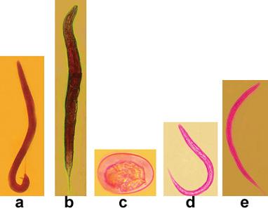
Figure (2): Microphotographs of different developmental stages and sexes of Strongyloides
stercoralis isolated from fresh stools samples.
(a) parasitic adult male ( stained with Safranin), magn‡. X64 ; (b) parasitic adult female, magn. X64; (c) egg, magn. X640; (d) rhabditiform larva, magn. X640; and (e) filariform larva, magn. X320. Pictures (b), (c), (d) and (e) were colored by a Computer Adobe Phot
oshop▪ CS. Each of these five pictures was transformed from its original magnified size to the resolution of 1200 pixels/inch with the quality of 12 (maximum) and large file compatible with A4 page format .
‡magn. stands for the term magnification that gives the value of how many times the actual size of the
specimen was magnified.
Eriso, Afro-Egypt J Infect Endem Dis 2014; 4(2): 69-79
Original article
DISCUSSION
strongyloidiasis. On the other hand, the larvae of S. stercoralis, recovered from fresh stools
The results obtained in this study project can
samples of those infected student children,
be defined as a set of achievement scored by
were practically observed moving actively
way of cross-sectional type of study. Standard
in the fields of vision under the objectives of
deviation of infection rate, in student children
compound light microscopes. With this truth in
mind, the student children who were positive
sanitation and economic status, was far
for S. stercoralis infection and did not
less (S1 =2.2%) than in those from poor
manifest affectedness with strongyloidiasis
environmental sanitation and economic status
immunocompetent.
(S2 =12.5%). Larger S2 indicated that the
infected children, adults and larvae of the
observed infection rate went up beyond the
parasite were confined to the digestive tract in
mean infection rate in the population of
which case the children were symptomless
student children. Student children from
and the S. stercoralis infection they had was
families of better economic status did live a
asymptomatic intestinal strongyloidiasis. In
they used to get water supply lines to wash
"gastropulmonary
strongyloidiasis"
their hands, clothes and bodies at their
"disseminated strongyloidiasis" stage in these
homes. Families might be in a better economic
infected participant student children.
position by having enough capital to carry out their own private business work in the central
Here it could be understood that the parasite
part of the city with better sanitation that
was silently hiding in the intestine of each of
would be comfortable to be hygienic and buy
the infected student children to develop to the
shoes for their student children that could not
lethal conditions of strongyloidiasis whenever
be afforded and done by poor families.
the immunity of the student child was broken
Parents who had educational skill and
down (weakened) by some risk factors. Such
government job were economically self-
a hidden pathogenic parasite was found out
sufficient so that they were able to buy shoes
from where it was hiding by carefully
for their student children, resulting in reduction
employing standard diagnostic procedures
in the infection rate with S. stercoralis. The
such as Baermann technique and displayed
fact that poor environmental sanitation and
with all its developmental stages and
poor economic income did form one of the
sexes. Hence, the parasitic males of S.
obvious causes for the increase of infection
stercoralis are present together with their
rates in the student children was evidenced by
parasitic females in the bodies of human hosts
the statistic of standard deviation and other
and this verified evidence is a spectacularly
targetful answer to the major question and objective of this study.
When the safranin stain was tried to stain the worms of S. stercoralis, for the first time, it
gave a very good dyeing effect. It stained the
worms red. Actually, safranin is well known
hermaphroditism) were the methods of
as the secondary stain (counter stain) applied
reproduction for S. stercoralis in human hosts
to the fixed preparations of bacteria. If the
as the parasitic males did not exist in human
bacteria are decolorized with alcohol, they
body [6]. S. stercoralis
will take up the safranin and appear red
parthenogenetic and can produce offspring
(gram-negative). If the bacterial cells are not
without being fertilized by the male. But the
decolorized , the safranin will have no effect
fact that parasitic males do exist can be
on the already stained preparation, and the
demonstrated in experimentally infected dogs
bacteria will remain blue or purple (gram-
[5]. In other words, this group of thought
positive) [23, 24].
stated that the adult female S. stercoralis is parthenogenetic & hermaphroditic in the
The student children who were positive for S.
mechanism of reproduction. No adult male S.
stercoralis infection were not revealing or not
stercoralis is known to exist, the adult female
manifesting affectedness with the disease,
is considered as being parthenogenetic [26].
being active in their daily lives like other
Another division of thought had concluded
that asexual reproduction was the method for
Eriso, Afro-Egypt J Infect Endem Dis 2014; 4(2): 69-79
Original article
the parasitic females of S. stercoralis to
Applying efficient preventive measures
reproduce in human hosts for the very reason
and devising effective treatment under
that parasitic males did not exist in the body
clinical supervision against a pathogenic
of humans [8]. However, let us take that both
parasite depend on deep and detailed
parthenogenesis and asexual reproduction
understanding about the biology and life
have the same meaning for the method of
cycle of the parasite.
reproduction. Is there any evidence to
The term curved tail was used by
generalize that adult female S. stercoralis is
authors for the posterior body part of males
parthenogenetic and hermaphroditic? Each of
that belong to free living generation
these groups of thought did not have any trace
found in soil [4]. The term was not
of substantiated and persuasive scientific
inclusive and unfit to define the actual
proof to be accepted in science. This was so
taxonomic morphology of both free-living
and parasitic males of S. stercoralis.
stercoralis with spicules everted out of their
The degree (extent) of being curved in the
spicule pouches were practically observed in
posterior body part of male S. stercoralis
fresh stools samples of participant student
is greatly variable among the male worms
children. The presence of many parasitic
of both free-living and parasitic ones in
males of S. stercoralis with everted spicules
a similar way in extent. The morphology
in fresh stools samples together with parasitic
of both parasitic and free living males is
females was a very strong evidence for the
the same. This was verified by growing
fact that there was copulation & fertilization.
free-living males in autoclaved topsoil that
Everted spicules of males are seen only at
was inoculated with fresh stools sample
times of mating.
obtained from an infected child before giving him treatment and incubated at
CONCLUSION
28ºC. When the morphology, including the
The result of this study had identified the
variation in the degree of curvature or
concepts of both parthenogenesis/asexual
coiling of the posterior body part, of these
reproduction and protandrogony, in the
free-living males was compared with that
parasitic generation of S. stercoralis, to be
of parasitic males, it was found to be
similar in both free living and parasitic
evidences, on the reproduction of S.
ascertained to be correct to differentiate
stercoralis in the parasitic generation in
and free-living males
human hosts that had been reacted to by
their respective
this paper could not be denied because they
stercoralis was a ventrally "curved or
were reported straight forward by authors
coiled posterior body part" in the males of
in modern textbooks, journals, and on the
this very parasite whereas that of the
internet. Due to those reports, in all
females was straight.
modern human parasitology textbooks,
In the life cycle of parasitic generation of
journal, and on the internet, the males of S.
S. stercoralis both parasitic male & female
stercoralis had been excluded (omitted)
must be included just like the free-living
from the life cycle of its parasitic
male & female in their life cycle.
generation in human hosts. It was possible for copulation to take place between the
This article is a realistic response to a
parasitic males and females to result in
chronic global problem that has remained
fertilization in the lumen of the human
unsolved for generations of man until now
host's gut and then the fertilized parasitic
and needs world-wide attention of human
female could burrow into the intestinal
parasitologists.
mucosa to lay eggs that would hatch soon. It is just like a domestic cock and a hen
Funding:
where it is the hen which goes to a nest
The financial support, covering the cost of
after mating to lay and incubate eggs and
children and technical assistance by assigning a medical doctor for prescription and clinical
Eriso, Afro-Egypt J Infect Endem Dis 2014; 4(2): 69-79
Original article
supervision were given by the Ethiopian
REFERENCES
Catholic Church (Dilla Don Bosco).
1. Mitreva M, McCarter JP, Martin J, Dante M,
Conflict of interest:
Wylie T, Chiapelli B, et al. Comparative genomics of gene expression in the parasitic
I confirm that I don't have any competitive
and free-living nematodes Strongyloides
conflict of interest with any body.
stercoralis and Caenorhabditis elegans. Genome Res [serial on the Internet]. 2004
Ethical approval:
[cited 2012 Oct.24]14: [about 6p.]. Available from:http://genome.cship.org/content/14/2/209. full
Ethical permission/clearance to perform the
2. Concha R, Harrington WJR, Rogers AI.
research work for the well-being of human
strongyloidiasis:
subjects was obtained from:- Dilla University,
management, and determinants of outcome. J
the Office of Gedio-Zone Administration, and
Clin Gastroenterol. 2005; 39: 203-11.
the Directors of the schools involved in the
3. Hunter CJ, Petrosyan M, Asch M. Dissemination of
study. The demand for the continuity of this
Strongyloides stercoralis in a patient with
study project and participation by the
systemic lupus erythematous after initiation of
participant student children and their parents
albendazole: a case report. J Med Case
was unusually high.
Reports [serial on the Internet]. 2008 May 14 [cited 2012 Oct. 24];2: [about 5p.]. Available from: http: //www.jmedicalcasereports.com/con
Don Bosco, i.e., Dilla Ethiopian Catholic
4. Gilles HM. Soil-transmitted helminths (geohel-
minths). In: Cook GC. editor. Manson's tropical
Church was very kind and quick to accept the
request forwarded by the researcher of this
ed., London: WB Saunders; 1996;
study for technical and medical assistance.
5. Paniker CKJ. Textbook of medical parasitology.
Contribution to and participation in this study
6th ed., New Delhi; Jaypee Brothers Medical
project by Don Bosco was immense. It was
Publishers 2007; 169-74.
this very organization that rescued the student
6. Bogitsh BJ, Carter CE, Oeltmann TN. Human
Strongyloides
parasitology. 3rd ed., USA; Academic press
stercoralis when it understood the potential
risk the infection could entail. Then it reacted
7. Dillard KJ, Saari SAM, Anttila M.
promptly by covering the financial cost of the
Strongyloides
stercorali
Finnish kennel. Acta Vet Scand [serial on the
drug of choice for treatment and assigned a
Internet]. 2007 Dec 12 [cited 2012 Oct 24]; 49:
medical doctor (Dr. Corazon B. Jaca FMA)
[about 5p.].Available from:
for prescription and clinical supervision in the
http://www.actavetscand.com/content/49/1/37
process of treatment. I am extremely thankful
8. Keiser PB, Nutman TB. Strongyloides stercoralis in
to Don Bosco for its extensive financial
the immunocompromised population. Clin
contribution and participation in favour of the
Microbiol Rev [serial on the Internet]. 2004 Jan
infected student children.
[cited 2012 Oct 24];17(1):[about 3p.]. Available from:http://www.ncbi.nlm.nih.gov>…>Clin
I am very grateful to the office of the
Microbiol Rev>v.17(1); Jan 2004
President of Dilla University and the main
Administrator Office of Gedio Zone for their
Hyperinfective strongyloidiasis: S. stercoralis
writing letters to the Directors of elementary
undergoes autoinfective burst in neonatal
schools, requesting them to be cooperative for
gerbils. J Parasitol 1999; 85: 286-9.
my research activities with their students.
10. Dionisio D, Manneschi LL, Lollo SD, Orsi A,
Tani A, Esperti F. Strongyloides stercoralis:
I am very strongly thankful to Mr. Tilahun
ultrostructural study of newly hatched larvae
Kebede, Mr. Mingizem Birhan, and Mr.
within human duodenal mucosa. J Clin Pathol
Tsegaye Hylu who are computer science
2000; 53: 110-6.
teachers & technicians in Dilla University, for
11. Massey HCJ, Nishi M, Chaudhary K, Pakpout
their providing me with and installing
N, Lok JB. Structure and developmental
technical computer tools such as Equation
Strongyloides
stercoralis.
Editor and Adobe Photoshop▪CS that were
Int J Parasitol 2003; 33: 1537-44.
essential in processing the data.
Eriso, Afro-Egypt J Infect Endem Dis 2014; 4(2): 69-79
Original article
12. Garg PK, Perry S, Dorn M, Hardcastle L,
19. Fardet L, Genereau T, Poirot JL, Guidet B,
Parsonnet J. Risk of intestinal helminth and
Kettaneh A, Cabane L. Severe strongyloidiasis
protozoan infection in a refugee population. Am
in corticosteroid-treated patients: case series
J Trop Med Hyg [serial on the Internet]. 2005
and literature review. J Infect 2007; 54:18-27.
Mar 20 [cited 2012 Oct 24]; 83(3): [about 4 p.].
20. Kerepesi LA, Hess JA, Leon O, Nolan TJ,
Available from: http: //www.ajtm.org/content/
Schad GA, Abraham D. Toll-like receptor
4(TLR4) is required for protective immunity to
13. Siddiqui AA, Berk SL. Diagnosis of Strongyloides
larval Strongyloides stercoralis in mice.
stercoralis. In: Ericsson CD, Steffen R, editors.
Microb Infect 2007; 9: 28-34.
Clin Infect Dis [serial on the Internet]. 2012
21. Rahif RH, Emara GY, Al-saqur IM. Epidemio-
Oct 15 [cited 2012 Oct 24]; 55(8): [about
logical study of intestinal nematodes in dogs in
8 p.]. Available from: http: //cid.oxfordjournals.
Basrah(Iraq): prevalence of helminthic ova in
org/content/33/7/ 1040.full
the fecal deposits. Iraqi J Vet Sci 2002; 16: 71-
14. Siegel MO, Simon GL. Is human immuno-
deficiency virus infection a risk factor for
22. Cheesbrough M. District laboratory practice in
Strongyloides stercoralis hyperinfection and
tropical countries. Part 1. India; Cambridge
dissemination? PLoS Negl Trop Dis [serial on
University Press 1998; 214-5.
the Internet]. 2012 Jul 31 [cited 2012 Oct 24];
23. Merck KGA. Microscopy: Safranin 0 (C.I.
6(7): [about 4 p.].Available from: http:// www.
50240) Certistain. [serial on the Internet]. 2011;
ncbi. nlm.nih.gov>…>PLoS Negl Trop Dis>
[about 1 p]. Available from:
v.6(7); Jul 2012
15. Ramanathan R, Nutman TB. Strongyloides
24. Boyd PF. Basic medical microbiology. 5th ed.
stercoralis
USA: Little, Brown and Co. (Inc). 1995; 557.
compromised Host. Japan J Parasitol [serial on
25. Beadle GW, Emerson R, Whitaker DM,
the Internet]. 2008 May [cited 2012 Oct. 24];
Lawson CA, Lewis RW, Biirmester MA et al.
10(2):[about 4 p.]. Available from: http://www.
Full text of "Animal tissue techniques". [serial
on the Internet]. USA: W.H. Freeman and Co.
16. Poonam P, Jayshree RS, Acharya RS, Hema S,
1962; [about 1]. Available from: http://archive.
Govind B, Suresh TM. Fulminant fatal
org/stream/ animaltissuech00hurr
Strongyloides stercoralis infection in a
26. Zeibig EA. Clinical parasitology: a practical
postchemo-therapy immuno-suppressed cancer
approach. London; W.B Saunders Co. 1997; 148.
patient. Med Pediatric Oncol 1999; 33: 504-5.
17. Jayshree RS, Hema S, Govind B, Suresh TM.
Peer reviewer: Samia Etewa: Professor of
Strongyloides
stercoralis
Parasitology, Faculty of Medicine, Zagazig
the stools during anticancer therapy. Indian J
University, Egypt.
Gastroenterol 2001; 20: 160- 1.
Editor: Tarik Zaher: Professor of Tropical
18. Kim J, Joo HS, Ko HM, Na MS, Hw ASH, Im
JC. A case of fatal hyperinfective strongyloidiasis
University, Egypt.
with discovery of autoinfective filariform larvae in sputum. Korean J Parasitol 2005; 43: 51-5.
Eriso, Afro-Egypt J Infect Endem Dis 2014; 4(2): 69-79
Source: http://mis.zu.edu.eg/ajied/Ajied_System_Files/p123.pdf
CorSalud 2014 Jan-Mar;6(1):36-46 Cuban Society of Cardiology Original Article Clinical and angiographic assessment of coronary bare-metal stent restenosis Max G. Sánchez Manzanaresa MD; Francisco L. Moreno-Martínezb, MD, MSc; Iguer F. Aladro Mirandab, MD, MSc; Luis F. Vega Fleitesb, MD; Rosendo S. Ibargollín Hernándezb,
The Right to Parenthood An Argument for a Narrow Interpretation1 ABSTRACTThe paper argues for two kinds of limitations on the right to parenthood. First, it claims that the right to parenthooddoes not entail a right to have as many children as one desires. This conclusion follows from the standard justificationsfor the right to parenthood, none of which establishes the need to grant special protection to having as many childrenas one desires. Second, with respect to the right to receive assistance from the state in IVF, it is suggested that the stateshould also be allowed to take non-medical considerations into account in determining whether or not an applicant isentitled to this service, particularly in cases where the applicant seems to lack mothering ability.