Jimd4221 681.693

J. Inherit. Metab. Dis. 25 (2002) 681 693# SSIEM and Kluwer Academic Publishers. Printed in the Netherlands.
The osteodystrophy of mucolipidosis type IIIand the effects of intravenous pamidronatetreatment OBINSON , N. BAKER , J. NOBLE , A. KING , G. DAVID , ILLENCE , P. HOFMAN and T. CUNDY * 1Department of Paediatrics, Nelson Hospital, Nelson; 2Department of Pathology,Middlemore Hospital, Auckland, New Zealand; 3Academic Department of MedicalGenetics, The Children's Hospital, Westmead, Australia; 4Faculty of Medicine &Health Sciences, University of Auckland, Auckland, New Zealand *Correspondence: Department of Medicine, Faculty of Medicine & Health Sciences,University of Auckland, Private Bag 92019, Auckland 1, New Zealand.
E-mail: [email protected] MS received 14.10.02 Accepted 15.11.02 Summary: Mucolipidosis type III (ML III; McKusick 252600) is a rare lysosomalstorage disease in which skeletal involvement is prominent, in particular thedestruction of vertebral bodies and the femoral heads. We describe studies intwo siblings with ML III that suggest the presence of a distinct metabolic bonedisorder. Biochemical indices of bone turnover were increased, and transiliacbone biopsy demonstrated both trabecular osteopenia and marked subperiostealbone resorption. Intravenous pamidronate treatment given monthly for a yearwas well tolerated and produced dramatic clinical effects, with reduction in bonepain and improvements in mobility, despite incomplete suppression of boneresorption as assessed by biochemical, radiographic and histological criteria.
Bisphosphonate therapy may have an important role in the management of bonepain in ML III, as it does in the related lysosomal disorder of Gaucher disease.
Mucolipidosis type III (McKusick 252600, also known as pseudo-Hurlerpolydystrophy) is a recessively inherited lysosomal storage disease. These disordersarise from defects in lysosomal enzymes responsible for the breakdown of complexmolecules, such as glycosaminoglycans. Tissue damage develops as a result of theaccumulation of incompletely processed macromolecules. The differing clinicalphenotypes of the lysosomal storage disorders reflect the enzyme that is defective,the products that accumulate, and the particular tissues in which they accumulate.
In the case of mucolipidosis type III, which was first described by Maroteaux and Lamy (1966), the defective enzyme is N-acetylglucosaminyl 1-phospho-transferase. This enzyme is important in the posttranslational modification of severallysosomal enzymes. It adds a mannose 6-phosphate recognition marker that permitsthe enzyme to be transported into lysosomes (and, to a lesser extent, permits thereuptake of enzyme into lysosomes from the extracellular milieu). Within thelysosome, further posttranslational modification of the enzymes takes place andthe acid environment is necessary for enzymatic function. In the absence of themannose 6-phosphate marker, the enzymes are not taken up into lysosomes andreadily ‘leak' out of cells. The plasma activities of these enzymes may thus be high,but they are ineffective (Kornfeld and Sly 2001; Reitman et al 1981). Unprocessedglycoproteins and glycolipids accumulate in tissues, giving rise to the clinical mani-festations of the disease.
The clinical manifestations of mucolipidosis type III (ML III) include a mild, Hurler-type dystrophy, mild intellectual disability, valvular heart disease andcorneal opacities. Orthopaedic and skeletal manifestations are well recognized(Haddad et al 2000; Hetherington et al 1999; Kornfeld and Sly 2001; Melhemet al 1973). These include the constellation of skeletal abnormalities common tothe mucopolysaccharidoses known as dysostosis multiplex, and also a progressivehip dysplasia and vertebral abnormalities (Table 1). In this paper we describe twosiblings with severe skeletal complications of ML III. Our observations suggest thatML III is characterized by a distinctive high-turnover metabolic bone disorder. These¢ndings, together with reports of bene¢cial e¡ects of bisphosphonates in anotherlysosomal storage disorder, Gaucher disease (Cianna et al 1997; Harinck et al 1984;Ostlere et al 1991; Samuel et al 1994), and the absence of any speci¢c treatmentfor either ML III or its bony sequelae, led us to trial intravenous pamidronate inthe siblings.
Orthopaedic and skeletal complications of mucolipidosis type III Sti¡ness of hands and shouldersClaw hand deformityScoliosisShort statureLow iliac wings, shallow acetabular fossaErosion of the femoral headsValgus deformity of femoral neckUnderdevelopment of posterior elements of dorsal spineDysostosis multiplex Skull: calvarial thickening, premature suture closure, shallow orbits, abnormal toothspacing, J-shaped sella Spine: anterior hypoplasia of lumbar vertebrae, kyphosis Long bones: shortened, wide diaphyses, irregular metaphyses, poorly developedepiphyseal centres Clavicles: short, thickened, irregular Ribs: narrow at vertebral ends, £at and broad at sternal ends Phalanges: shortened, trapezoidal in shape, widened diaphyses J. Inherit. Metab. Dis. 25 (2002) Osteodystrophy of mucolipidosis III This boy was diagnosed at 6 years of age following investigations for symptoms of joint pain and sti¡ness. The diagnosis of ML III was con¢rmed onthe basis of measurements of lysosomal enzyme activities in plasma and culturedskin ¢broblasts (Table 2). Although he had symptoms of joint pain and restrictedmovement for many years, he remained mobile until adolescence. At the age of15 years he developed progressive walking di⁄culties. Magnetic resonance imagingshowed an expansion of the extradural tissue anteriorly opposite C2 C7. A cervicaldecompression was performed, with fusion at C7, but his neurological status con-tinued to deteriorate. He has been e¡ectively paraplegic and wheelchair-bound since.
The reason he failed to improve following surgery is not certain. Possiblemechanisms include cord compression, intramedullary storage, vertebral collapseor central nervous system degeneration as a result of lysosomal storage. At 18 yearsof age, subject A was su¡ering from signi¢cant hip and back pain, which wasseriously compromising his quality of life, waking him from sleep and interfering withwheelchair transfers.
Subject B is the younger sister of subject A. She was diagnosed with ML III at the age of 2 years following her brother's diagnosis (Table 2). By the age of 12 yearsshe was also su¡ering from signi¢cant, constant joint pain, mainly from the hips. Thiswas accompanied by a dramatic deterioration in her mobility. By age 14 years shewas barely able to walk 5 metres and was spending most of her time in a wheelchair.
Both subjects A and B had tried several forms of oral analgesia in an attempt to decrease their hip and back pain. These had all been unsuccessful. In light of experi-ence suggesting bene¢cial e¡ects of bisphosphonates in relieving bone pain inosteopenic disorders (Astrom and Soderhall 1998; Brumsen et al 1997; Glorieuxet al 1998; Landsmeer-Beker et al 1997; Zacharin and Cundy 2000), ¢brous dys-plasia (Chapurlat et al 1997; Lala et al 2000) and Gaucher disease (Cianna et al 1997;Harinck et al 1984; Ostlere et al 1991; Samuel et al 1994), we elected to treat bothsubjects with pamidronate, a second-generation aminobisphosphonate.
Lysosomal enzyme activity was measured in blood samples (in both plasma andleukocytes), and in cultured skin fibroblasts (in both cells and the supernatant), bythe Australian National Lysosomal Diseases Reference Laboratory (Women's andChildren's Hospital, Adelaide, South Australia). Total plasma alkaline phosphataseactivity and plasma osteocalcin (Nichols Diagnostics, San Clemente CA, USA) wereused as the main markers of bone formation. The fasting urine ratios ofhydroxyproline/creatinine and N-telopeptide/creatine (breakdown products of typeI collagen) were used as markers of bone resorption. Urinary N-telopeptide wasmeasured using the Osteomark NTx assay (Ostex International Inc., Seattle, WA, USA).
Bone age was determined from hand radiographs by the method of Tanner and Whitehouse (1983). The combined cortical width of the second metacarpal was J. Inherit. Metab. Dis. 25 (2002) NRa In are enzym high J. Inherit. Metab. Dis. 25 (2002) Osteodystrophy of mucolipidosis III measured at its midpoint, using a dial-gauge micrometer (by subtracting the medullarywidth from the total width).
Bone mineral content (BMC, g), and bone area (BA, cm2) were measured by dual x-ray absorptiometry (DEXA, Lunar DPX-L, Madison, WI, USA). Measurementswere made at the lumbar spine in subject B, and at the forearm in subject A (spinalmeasurements were not possible in subject A because of deformity and mobilitydi⁄culties). From the lumbar spine measurements, the derived values of areal bonemineral (vBMD ¼ aBMD/ BA, g/cm3) were calculated. Forearm measurements of BMC,bone width and the aBMD of a 1.5 cm length segment were made at both theultradistal radius and ulna sites (predominantly trabecular bone), and the samemeasurements were made on a 2 cm length segment at both the 33% radius and ulnasites (predominantly cortical bone). Areal BMD values were also expressed as anage- and sex-matched standard deviation score (z-score) according to themanufacturer's database.
Transiliac bone biopsies (8 mm trephine) were taken before and after one year of month- ly pamidronate infusions. Undecalci¢ed sections were stained with von Kossa andGoldner's trichrome stain. Quantitative histomorphometry was undertaken using theOsteoMeasure system (Atlanta, GA, USA). The specimens were not tetracycline labelled.
Both subjects received monthly intravenous infusions of pamidronate: the initial dose given was 7.5 mg/m2 body surface area, and subsequent doses were 15 mg/m2,infused in 150 ml of saline over 4 h. Calcitriol, 0.25 mg twice a day and a daily oralcalcium supplement were given for a week after each intravenous treatment.
Laboratory ¢ndings: Before treatment, serum calcium, phosphate and parathyroid hormone concentrations remained within age- and sex-speci¢c reference ranges. Renalfunction remained normal. Plasma alkaline phosphatase activity and plasmaosteocalcin concentrations were increased in both subjects before pamidronatetreatment, as was the fasting urine hydroxyproline/creatinine ratio (Table 3).
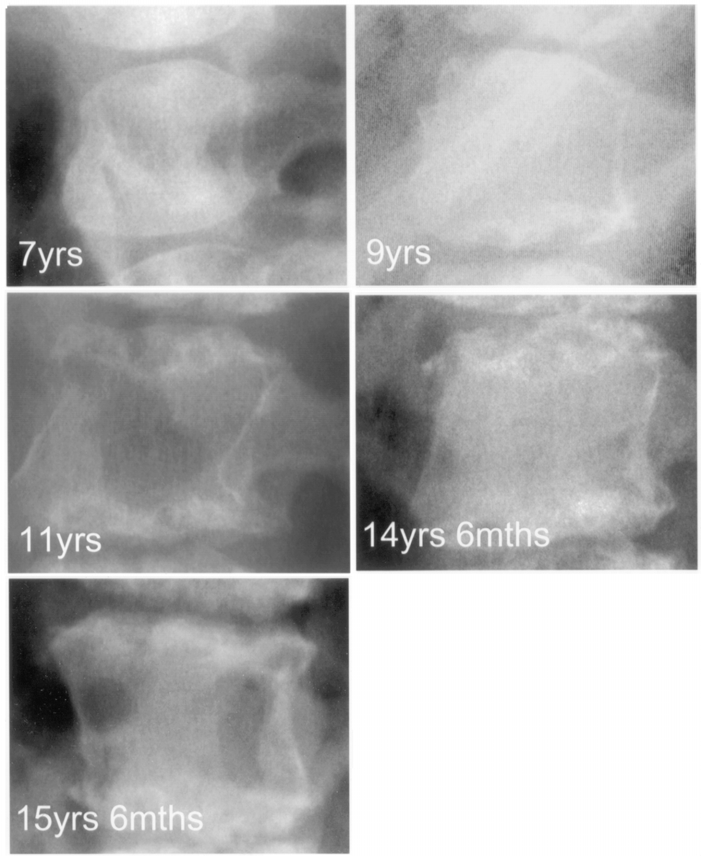
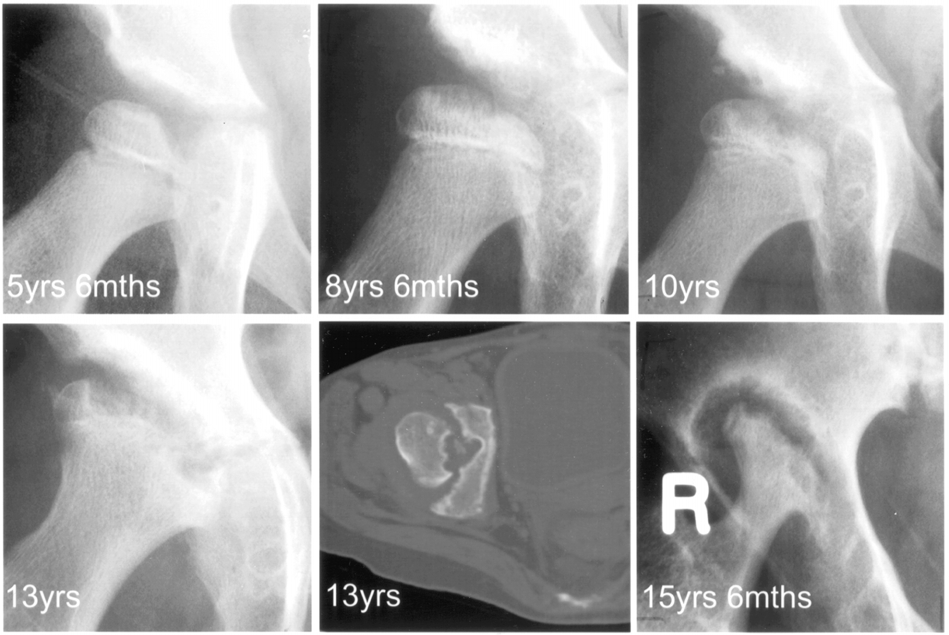
Radiology and bone denistometry: Skeletal radiographs showed typical changes of dysostosis multiplex but also progressive erosive destruction of the hip joints andthe superior endplates of the vertebrae (Figures 1 and 2). Both subjects had delayedskeletal maturation. At the time pamidronate treatment was started, the bone ageof subject A was 17.2 years (chronological age 18.9), and the bone age of subjectB was 12.9 years (chronological age 14.6).
Bone densitometry at the forearm in subject A showed reduced z-scores of 3.7 at the ultradistal radius (trabecular) site and 3.5 at the proximal radius (cortical) site.
In subject B the z-scores were 3.1 at the lumbar spine (predominantly trabecularbone) and 2.1 for the whole body scan (predominantly cortical bone). These valuesare somewhat di⁄cult to interpret in view of the short stature of both subjects.
The estimated volumetric density of the lumbar spine in subject B was 0.278 g/cm3,which is close to the mean for this age.
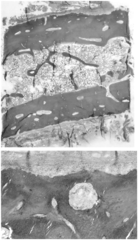
J. Inherit. Metab. Dis. 25 (2002) Biochemical indices of bone turnover before and during pamidronate therapy Alkaline phosphatase (iu/L) Osteocalcin (mg/L) (mmol/mmol creatinine) (nmol BCE/mmol creatinine) aNormal values appropriate for age and sex The ¢ndings in both subjects were similar. The most striking ¢nding was of vigorous osteoclastic subperiosteal bone resorption occupying almostall the periosteal surface (Figure 3). There was evidence of active endosteal modellingwith occasional osteoclasts and slight marrow ¢brosis. Osteoid surfaces wereincreased in extent but were of normal thickness. The cortical widths were normal,but trabecular bone volumes were low in both subjects"more than three standardsdeviations below age-expected values (Glorieux et al 2000).
E¡ects of pamidronate therapy: Cyclic intravenous pamidronate treatment was well tolerated, apart from ¢rst-dose febrile responses, necessitating the admission of sub-ject B to hospital for one night. Both subjects had a dramatic clinical response topamidronate treatment, with both decreased pain and improved mobility. SubjectA became almost completely pain free and able to assist in his wheelchair transfers.
Subject B became pain free and had markedly improved mobility, being able touse crutches or a walking frame for moving about the house and garden, but stillneeding her wheelchair for longer trips. Both the patients and their parents reporteda dramatic improvement in quality of life, including better sleep. Pubertal develop-ment in subject B proceeded normally, with menarche occurring a year after startingpamidronate.
No signi¢cant change in plasma calcium, phosphate or parathyroid hormone occurred during treatment. Markers of bone turnover were increased at baselinein both subjects, more so in subject B, whose growth was not yet complete. Somesuppression of both urine hydroxyproline excretion and plasma alkaline phosphataseactivity occurred during the ¢rst year's treatment with pamidronate, but in neithersubject were normal values reached. Measurements of N-telopeptide/creatinineexcretion made at the end of this period con¢rmed that bone resorption remainedhigh (Table 3).
Repeat skeletal surveys taken after one year's pamidronate therapy showed increased bone density, particularly in the metaphyseal regions. The density of cortical J. Inherit. Metab. Dis. 25 (2002)


Osteodystrophy of mucolipidosis III 1 = 25 pamidronate J. Inherit. Metab. Dis. 25 (2002)


Lateral radiographs of the ¢rst lumbar vertebra of subject B, at ages 7, 9, 11, 141=2, and 151=2 years, showing the appearance of dysostosis multiplex in the early ¢lms, with anteriorhypoplasia of the vertebral body, but also progressive erosion of the superior and inferiorendplates. The ¢nal picture, obtained after one year's pamidronate therapy, shows increasedradiographic density at the end plates, but persistence of the erosive changes. The appearanceswere similar in subject A J. Inherit. Metab. Dis. 25 (2002)


Osteodystrophy of mucolipidosis III Bone histology from subject B, taken before pamidronate treatment. Goldner stain.
The low-power view (top) demonstrates the very low trabecular bone volume (11%, more than3 standard deviations below normal). On the high-power view (bottom), extensive osteoclasticresorption is visible all along the periosteal surface bone also appeared to increase after pamidronate. The combined cortical width at themidpoint of the second metacarpal decreased by 7% in subject A and increased by 10%in subject B. The radiographic density of the femoral heads, acetabuli and vertebralendplates had increased, but the erosive changes had not changed signi¢cantly(Figures 1 and 2).
J. Inherit. Metab. Dis. 25 (2002) Bone densitometry was repeated 6-monthly. In subject A the BMC at the ultradistal radius and ulna sites (trabecular bone) showed increases of 50% and 76%, respectively,over one year, without change in bone width. At the proximal radius and ulna sites(cortical bone), BMC increased 18% and 29%, respectively, again with no changein bone width. At the ultradistal radius site, the areal bone density z-score increasedfrom 3.7 to 0.1. At the proximal radius site, it increased from 3.5 to 2.5.
In subject B the BMC of the 2nd 4th lumbar vertebrae increased by 70%, an e¡ectpartly arising from a 31% increase in estimated bone volume and partly from a29% increase in estimated volumetric bone density. The areal bone density z-scoreincreased from 3.1 to 1.4.
Repeat bone biopsies after one year's therapy with pamidronate showed an essentially unchanged picture with vigorous subperiosteal bone resorption.
The two siblings that we studied both demonstrated the characteristic severe erosivebone disorder of ML III (Haddad et al 2000; Hetherington et al 1999; Kornfeldand Sly 2001; Melhem et al 1973), particularly affecting the femoral heads, acetabulaeand vertebrae. The bone disorder was first evident radiographically around the age of10 years and progressed relentlessly. The biochemical data indicate that both boneresorption and bone formation were increased, suggesting a ‘‘metabolic'' bone dis-order. The higher values of bone turnover markers in the younger sibling probablyreflect the fact that her growth was less complete. The pathology of bone in MLIII has not been reported before. The finding of marked bone resorption at the per-iosteal surface is quite distinctive. Similar findings were noted in the only publishedreport of bone histology in the closely related disorder of mucolipidosis II (Pazzagliaet al 1989). In that case, the similarity with the histological findings in osteitis fibrosacystica led to the suggestion that hyperparathyroidism might be responsible for thesubperiosteal bone resorption. However, the normal pretreatment plasma concen-trations of calcium, phosphate and parathyroid hormone in our subjects make thisimplausible. Others have suggested that the changes at the femoral head representosteonecrosis (Haddad et al 2000), but a recent study of the MRI appearances ofthe hip in ML III did not show characteristic features of osteonecrosis (Wihlborget al 2000).
The ¢nding of accelerated bone resorption is somewhat surprising. Although most of the lysosomal enzymes of osteoclasts are destined for secretion (rather than anintracellular function), mannose 6-phosphate signalling does appear to be importantin osteoclasts. Mannose 6-phosphate (M-6-P) receptors are found in the endoplasmicreticulum, in the Golgi stacks, and in transport vesicles that fuse with the ru¥edborder membrane. M-6-P signalling appears to be involved in the tra⁄cking ofosteoclastic lysosomal enzymes to the apical membrane of the osteoclast, where theyare secreted into the sealed, bone-resorbing compartment (Baron et al 1988).
Thus, one might expect osteoclast activity to be reduced when there is defective M-6-Pdirection of the bone-resorbing enzymes. However, osteoclastic enzymes are meant tobe secreted, so it is possible that, through leakage, greater than normal concentrations J. Inherit. Metab. Dis. 25 (2002) Osteodystrophy of mucolipidosis III accumulate within the sealed zone. Furthermore, not all bone-resorbing osteoclasticenzymes have M-6-P signalling. Acid phosphatase, for example, does not. It issynthesized as a transmembrane protein that is proteolysed to a soluble form onarrival in the lysosome, and is targeted to lysosomes by a pathway that does notrequire M-6-P (Barriocanal et al 1986; Kornfeld and Sly 2001). In the enzyme studieson cultured ¢broblasts (Table 2), it is interesting to note that acid phosphatase activitywas increased in the intracellular, as well as the extracellular, compartments, perhapssuggesting an increase in acid phosphatase synthesis.
Bisphosphonates are synthetic analogues of pyrophosphate that bind to the hydroxyapatite lattice of bone and are incorporated into the skeletal matrix. Theyare antiresorptive, an e¡ect that is mediated via suppression of osteoclast numbersand function. The aminobisphosphonates are taken up by osteoclasts and incorpor-ated into phosphorylated compounds within the mevalonate metabolic pathway. Thisinterferes with the synthesis of geranylgeranyl diphosphate and fumaryl diphosphate,which in turn interferes with the membrane localization of small GTPases (Ras, Rhoand Rac). These are important signalling proteins involved in transport processes inthe osteoclast and also with isoprenylation of transported proteins (Rogers et al 2000).
The ¢rst description of bisphosphonate use in children was in 1969, in an attempt to treat myositis ossi¢cans progressiva (Bassett et al 1969). In recent yearsbisphosphonates have been used safely, and with success, in children in a numberof conditions including hypercalcaemia, ¢brous dysplasia, and various osteoporoticdisorders (Shoemaker 1999). A commonly reported ¢nding in many of these studiesof bisphosphonates in children is the marked analgesic e¡ect on bone pain, whichis often reported by patients and their caregivers early in the course of treatment.
This phenomenon was certainly very noticeable in the subjects we studied. The causeof bone pain is uncertain, and the reason why bisphosphonates are e¡ective is evenmore so. There are examples where the analgesic e¡ects of bisphosphonates seemto be relatively dissociated from other e¡ects. In both Gaucher disease and ¢brousdysplasia, remarkable e¡ects on bone pain can be observed with relatively little changein extent of the pathological lesions (Chapurlat et al 1997; Cianna et al 1997; Harincket al 1984; Lala et al 2000; Ostlere et al 1991; Samuel et al 1994). In the subjects withML III that we studied, it was notable that we obtained incomplete suppressionof bone resorption as assessed by hydroxyproline and N-telopeptide excretion,and by bone histology. This is at variance with the ¢ndings in other types of paediatricbone diseases. We have subsequently used higher doses of intravenous pamidronate inour patients and still failed to suppress these markers of bone resorption (data notshown). The bone density data indicate that pamidronate did produce substantiale¡ectsPwith increases in bone density particularly in trabecular bone-rich areas.
Why bone resorption on the periosteal surfaces is so active, and why it is so di⁄cultto suppress, are intriguing but currently unanswered questions.
In conclusion, we have presented evidence for the existence of a distinctive osteodystrophy in ML III. The use of intravenous pamidronate was associated withremarkable symptomatic improvement in bone pain, analgesic requirements andmobility, despite incomplete suppression of the abnormal bone turnover. Pamidronatetherapy was well tolerated and safe.
J. Inherit. Metab. Dis. 25 (2002) We wish to thank Roland Baron for his helpful comments, and to acknowledge theassistance of the National Referral Laboratory for Lysosomal, Peroxisomal andRelated Genetic Disorders, North Adelaide, South Australia for the diagnosticstudies. Grace David is funded by a grant from the ConnecTed committee, Australia.
Astrom E, Soderhall S (1998) Bene¢cial e¡ect of bisphosphonate during ¢ve years of treatment of severe osteogenesis imperfecta. Acta Paediatr 87: 64 68.
Bassett CAL, Donath A, Macagno F, Preisig R, Fleisch H, Francis MD (1969) Diphosphonates in the treatment of myositis ossi¢cans. Lancet ii: 845.
Baron R, Ne¡ L, Brown W, Courtoy PJ, Louvard D, Farquhar MG (1988) Polarized secretion of lysosomal enzymes: co-distribution of cation-independent mannose-6-phosphate receptorsand lysosomal enzymes along the exocytic pathway. J C ell Biol 106: 1863 1872.
Barriocanal JG, Bonifacino JS, Yuan L, Sandoval IV (1986) Biosynthesis, glycosylation, movement through the Golgi system, and transport to lysosomes by an N-linkedcarbohydrate-independent mechanism of three lysosomal integral membrane proteins.
J Biol C hem 261: 16755 16763.
Brumsen C, Hamdy NA, Papapoulos SE (1997) Long-term e¡ects of bisphosphonates on the growing skeleton. Studies of young patients with severe osteoporosis. Medicine 76: 266 283.
Chapurlat RD, Delmas PD, Liens D, Meunier PJ (1997) Long-term e¡ects of intravenous pamidronate in ¢brous dysplasia of bone. J Bone Miner Res 12: 1746 1752.
Cianna G, Cuttini M, Bembi B (1997) Short-term e¡ects of pamidronate in patients with Gaucher's disease and severe skeletal involvement. N Engl J Med 337: 712.
Glorieux FH, Bishop NJ, Plotkin H, Chabot G, Lanoue G, Travers R (1998) Cyclic admin- istration of pamidronate in children with severe osteogenesis imperfecta. N Engl J Med339: 947 952.
Glorieux FH, Travers R, Taylor A, et al (2000) Normative data for iliac bone his- tomorphometry in growing children. Bone 26: 103 109.
Haddad FS, Hill RA, Vellodi A (2000) Orthopaedic manifestations of mucolipidosis type III: an illustrative case. J Pediatr Orthop 9: 58 61.
Harinck HI, Bijvoet OL, Van der Meer JW, Jones B, Onvlee GJ (1984) Regression of bone lesions in Gaucher's disease during treatment with aminohydroxypropylidenebisphosphonate. Lancet ii: 513.
Hetherington C, Harris NJ, Smith TWD (1999) Orthopaedic management in four cases of mucolipidosis type III. J R Soc Med 92: 244 246.
Kornfeld S, Sly WS (2001) I-cell disease and pseudo-Hurler polydystrophy: disorders of lysosomal enzyme phosphorylation and localization. In: Scriver CR, Beaudet AL, SlyWS, Valle D, eds; Childs B, Kinzler KW, Vogelstein B, assoc. eds. The Metabolic and Mol-ecular Bases of Inherited Disease, 8th edn, vol III. New York: McGraw-Hill, 3469 3482.
Lala R, Matarazzo P, Bertelloni S, Buzi F, Rigon F, de Sanctis C (2000) Pamidronate treatment of bone ¢brous dysplasia in nine children with McCune Albright syndrome. Acta Paediatr89: 188 193.
Landsmeer-Beker EA, Massa GG, Maaswinkel-Mooy PD, van de Kamp JJP, Papapoulos SE (1997) Treatment of osteogenesis imperfecta with the bisphosphonate olpadronate(dimethylaminohydroxypropylidene bisphosphonate). Eur J Pediatr 156: 792 794.
Maroteaux P, Lamy M (1996) La pseudo-polydystrophie de Hurler. Presse Med 74: 2889 2892.
Melhem R, Dorst JP, Scott CI, McKusick VA (1973) Roentgen ¢ndings in mucolipidosis III (pseudo-Hurler polydystrophy). Radiology 106: 153 160.
J. Inherit. Metab. Dis. 25 (2002) Osteodystrophy of mucolipidosis III Ostlere L, Warner T, Meunier PJ, et al (1991) Treatment of type 1 Gaucher's disease a¡ecting bone with aminohydroxypropylidene bisphosphonate (pamidronate) Q J Med 79: 503 515.
Pazzaglia UE, Belu⁄ G, Campbell JB, et al (1989) Mucolipidosis II: correlation between radiological features and histopathology of the bones. Pediatr Radiol 19: 406 413.
Reitman ML, Varki A, Kornfeld S (1981) Fibroblasts from patients with I-cell disease and pseudo-Hurler polydystrophy are de¢cient in uridine 50-diphosphate-N-acetylglucosamine:glycoprotein N-acetylglucoaminylphosphotransferase activity. J Clin Invest 67: 1574 1579.
Rogers MJ, Gordon S, Benford HL, et al (2000) Cellular and molecular mechanisms of action of bisphosphonates. Cancer 88 (supplement 12): 2961 2978.
Samuel R, Katz K, Papapoulos SE, Yosipovitch Z, Zaizov R, Leiberman UA (1994) Aminohydroxypropylidene bisphosphonate (APD) treatment improves the clinical skeletalmanifestations of Gaucher's disease. Pediatrics 94: 385 389.
Shoemaker LR (1999) Expanding role of bisphosphonate therapy in children. J Pediatr 134: 264 267.
Tanner JM, Whitehouse RH, Cameron N, Marshall WA, Healy MJR, Goldstein H (1983) Assessment of Skeletal Maturity and Prediction of Adult Height, 2nd edn. London.
Academic Press.
Wihlborg CEM, Babyn PS, Clarke JTR (2000) MRI appearances of hip abnormalities in mucolipidosis type III. Pediatr Radiol 30: 262 264.
Zacharin M, Cundy T (2000) Osteoporosis pseudoglioma syndrome: treatment of spinal osteoporosis with intravenous bisphosphonates. J Pediatr 137: 410 415.
J. Inherit. Metab. Dis. 25 (2002)

Source: http://www.postpolio.org.nz/__data/assets/pdf_file/0015/10374/Osteodystrophy_of_Mucolipidosis_Type_III.pdf

Slide

Phair, Yasmin S. and Miller, Paul K. (2015) Inhibition of pain during the rehabilitation of knee ligament injuries: an interpretive phenomenological analysis. In: BASES Student Conference, 31 March - 1 April 2015, Liverpool John Moore's University, UK. Usage of any items from the University of Cumbria Repository ‘Insight' must conform to the following fair usage guidelines:

selfhelp.on.ca

J Med Humanit (2014) 35:257–271DOI 10.1007/s10912-014-9293-5 The Icarus Project: A Counter Narrative for PsychicDiversity Sascha Altman DuBrul Published online: 17 July 2014# Springer Science+Business Media New York 2014 Abstract Over the past 12 years, I've had the good fortune of collaborating with others tocreate a project which challenges and complicates the dominant biopsychiatric model ofmental illness. The Icarus Project, founded in 2002, not only critiqued the terms and practicescentral to the biopsychiatric model, it also inspired a new language and a new community forpeople struggling with mental health issues in the 21st century. The Icarus Project believes thathumans are meaning makers, that meaning is created through developing intrapersonal andinterpersonal narratives, and that these narratives are important sites of creativity, struggle, andgrowth. The Icarus counter narrative and the community it fostered has been invaluable forpeople around the world dealing with psychic diversity—particularly for people alienated bymainstream approaches. But, despite the numbers of people who have been inspired by thisapproach, the historical background of the Icarus Project is hard to find. It exists primarily inoral history, newspaper articles, unpublished or self-published Icarus documents, and ininternet discussion forums. As the co-founder of the Icarus Project, I use this article to makemy understanding of that history and its key documents more widely available.

Copyright © 2008-2016 No Medical Care