Kamagra gibt es auch als Kautabletten, die sich schneller auflösen als normale Pillen. Manche Patienten empfinden das als angenehmer. Wer sich informieren will, findet Hinweise unter kamagra kautabletten.
V-cbc.ca
Supplemental Material can be found at:
0022-3565/10/3322-569–577$20.00THE JOURNAL OF PHARMACOLOGY AND EXPERIMENTAL THERAPEUTICS
Copyright 2010 by The American Society for Pharmacology and Experimental Therapeutics
JPET 332:569–577, 2010
Printed in U.S.A.
Cannabidiol Displays Antiepileptiform and AntiseizureProperties In Vitro and In Vivo□S
Nicholas A. Jones, Andrew J. Hill, Imogen Smith, Sarah A. Bevan, Claire M. Williams,Benjamin J. Whalley, and Gary J. Stephens
School of Pharmacy (N.A.J., A.J.H., I.S., S.A.B., B.J.W., G.J.S.) and School of Psychology (N.A.J., A.J.H., C.M.W.),University of Reading, Whiteknights, Reading, United Kingdom
Received July 22, 2009; accepted November 9, 2009
ABSTRACT
Plant-derived cannabinoids (phytocannabinoids) are com-
(in CA1, only at 100 M CBD), burst duration (in CA3 and
pounds with emerging therapeutic potential. Early studies
DG), and burst frequency (in all regions). CBD (1, 10, and 100
suggested that cannabidiol (CBD) has anticonvulsant prop-
mg/kg) effects were also examined in vivo using the penty-
erties in animal models and reduced seizure frequency in
lenetetrazole model of generalized seizures. CBD (100 mg/
limited human trials. Here, we examine the antiepileptiform
kg) exerted clear anticonvulsant effects with significant de-
and antiseizure potential of CBD using in vitro electrophys-
creases in incidence of severe seizures and mortality
iology and an in vivo animal seizure model, respectively. CBD
compared with vehicle-treated animals. Finally, CBD acted
(0.01–100 M) effects were assessed in vitro using the
with only low affinity at cannabinoid CB
Mg2⫹-free and 4-aminopyridine (4-AP) models of epilepti-
displayed no agonist activity in [35S]guanosine 5⬘-
O-(3-thio)-
form activity in hippocampal brain slices via multielectrode
triphosphate assays in cortical membranes. These findings
array recordings. In the Mg2⫹-free model, CBD decreased
suggest that CBD acts, potentially in a CB receptor-inde-
epileptiform local field potential (LFP) burst amplitude
pendent manner, to inhibit epileptiform activity in vitro and
[in CA1 and dentate gyrus (DG) regions] and burst duration
seizure severity in vivo. Thus, we demonstrate the potential
(in all regions) and increased burst frequency (in all regions).
of CBD as a novel antiepileptic drug in the unmet clinical
In the 4-AP model, CBD decreased LFP burst amplitude
need associated with generalized seizures.
A growing number of phytocannabinoids have been shown
endocannabinoid (eCB) system has been shown to be a key
to possess biological activity (Pertwee, 2008) and, in partic-
determinant of hippocampal epileptiform activity (Wallace et
ular, to affect neuronal excitability in the CNS. Phytocan-
al., 2002; Monory et al., 2006; Luda´nyi et al., 2008). The
nabinoid actions are reported to be mediated by G protein-
major psychoactive compound ⌬9-THC was the first phyto-
coupled cannabinoid CB and CB receptors and potentially
cannabinoid reported to affect epileptiform activity; ⌬9-THC,
by other non-CB receptor targets (Howlett et al., 2004; Per-
a partial agonist at CB
receptors, was shown to inhibit
twee, 2008). CB receptors are highly expressed in the hip-
excitatory glutamatergic neurotransmission in hippocampal
pocampus (Herkenham et al., 1990; Tsou et al., 1998) and are
neurons under low Mg2⫹ conditions (Shen and Thayer, 1999;
well known to modulate epileptiform and seizure activity
but see Straiker and Mackie, 2005).
(Shen and Thayer, 1999; Wallace et al., 2001). Moreover, the
CBD is the major nonpsychoactive component of
Cannabis
sativa whose structure was first described by Mechoulam
This work was supported by a GW Pharmaceuticals and Otsuka Pharma-
and Shvo (1963); CBD has recently attracted renewed inter-
ceuticals award, by a University of Reading Research Endowment Trust Fund
est for its therapeutic potential in a number of disease states
award; and The Wellcome Trust [Grant 070739].
Article, publication date, and citation information can be found at
(Pertwee, 2008). CBD has been proposed to possess anticon-
vulsive, neuroprotective, and anti-inflammatory properties
in humans. Thus, within the CNS, CBD has been proposed to
S The online version of this article (available at http://jpet.aspetjournals.org)
contains supplemental material.
be protective against epilepsy, anxiety, and psychosis and to
ABBREVIATIONS: CNS, central nervous system; CB, cannabinoid; eCB, endocannabinoid; ⌬9-THC, ⌬9-tetrahydrocannabinol; CBD, cannabidiol;
AED, antiepileptic drug; MEA, multielectrode array; aCSF, artificial cerebrospinal fluid; 4-AP, 4-aminopyridine; LFP, local field potential; NMDA,
N-methyl-D-aspartate; BSA, bovine serum albumin; SR141716A,
N-(piperidin-1-yl)-5-(4-chlorophenyl)-1-(2,4-dichlorophenyl)-4-methyl-1
H-
pyrazole-3-carboxamide; AM251,
N-(piperidin-1-yl)-5-(4-iodophenyl)-1-(2,4-dichlorophenyl)-4-methyl-1
H-pyrazole-3-carboxamide; GTP␥S,
guanosine 5⬘-
O-(3-thio)triphosphate; WIN55,212-2, [2,3-dihydro-5-methyl-3-(4-morpholinylmethyl)pyrrolo[1,2,3-
de]-1,4-benzoxazin-6-yl]-
1-napthalenylmethanone; PTZ, pentylenetetrazole.
Jones et al.
ameliorate diseases of the basal ganglia, such as parkinson-
binocular microscope (Leica AG, Solms, Germany). Dissected hip-
ism and Huntington's disease (Iuvone et al., 2009; Scuderi
pocampi were then adhered to the cleaned MEA surface using an
et al., 2009). CBD neuroprotective effects may be augmented
applied and evaporated cellulose nitrate solution in methanol (⬃4 l,
by reported antioxidant properties (Hampson et al., 1998;
0.24% w/v; Thermo Fisher Scientific, Leicestershire, UK) to ensure
Sagredo et al., 2007). Early studies suggested that CBD had
maximum contact between the tissue and recording electrodes and toavoid any physical stress on the tissue during recordings. Slices were
anticonvulsant potential in one small-scale phase I clinical
observed at 4⫻ magnification with a Nikon TS-51 inverted micro-
trial (Cunha et al., 1980). In this regard, there is a significant
scope (Nikon, Tokyo, Japan) and imaged via a Mikro-Okular camera
unmet clinical need for epilepsy, with ⬃30% of epileptic pa-
(Bresser, Rhede, Germany) to map electrode positions to hippocam-
tients experiencing intractable seizures regardless of conven-
pal regions. Slices were maintained at 25°C, continuously superfused
tional AED treatment (Kwan and Brodie, 2007). CBD is
(⬃2 ml/min) with carboxygenated aCSF, and allowed to stabilize for
extremely well tolerated in humans; for example, CBD at
at least 10 min before recordings. Signals were amplified (1200⫻
doses of 600 mg does not precipitate any of the psychotic
gain), band pass-filtered (2–3200 Hz) by a 60-channel amplifier
symptoms associated with ⌬9-THC (Bhattacharyya et al.,
(MEA60 System, Multi Channel Systems), and simultaneously sam-
2009). At present, CBD is used therapeutically in Sativex (1:1
pled at 10 kHz per channel on all 60 channels. Data were transferred
⌬9-THC/CBD; GW Pharmaceuticals, Porton Down, UK) to
to PC using MC_Rack software (Multi Channel Systems). Offlineanalysis of CBD effects upon burst amplitude, duration, and fre-
alleviate pain symptoms in multiple sclerosis and cancer
quency was performed using MC_Rack, MATLAB 7.0.4. (Mathworks
pain. CBD has anticonvulsant effects in animal models of
Inc., Natick, MA) and in-house analysis scripts. Animated contour
maximal electroshock (Karler et al., 1974; Consroe and
plots of MEA-wide neuronal activity (Supplemental Fig. 1) were
Wolkin, 1977; Consroe et al., 1982); however, CBD remains
constructed from raw data files processed in MATLAB 6.5 using
untested in other animal seizure models (Gordon and Devin-
in-house code with functions adapted from MEA Tools and interpo-
sky, 2001) and so has yet to fulfill its potential indications as
lated using a five-point Savitzky-Golay filter in MATLAB (Egert
a clinical anticonvulsant.
et al., 2002b). These data are displayed as peak source and peak sink
In the present study, we demonstrate the potential of CBD
animation frames. Burst propagation speeds were calculated by de-
as an AED. We show that CBD caused concentration-related
termining burst peak times at electrode positions closest to burst
and region-dependent attenuation of chemically induced ep-
initiation (CA3) and termination (CA1) sites using MC_Rack and
ileptiform activity in hippocampal brain slices using in vitro
ImageJ software (Abramoff et al., 2004).
Data Presentation and Statistics. Application of Mg2⫹-free and
MEA electrophysiological recordings. Furthermore, CBD re-
4-AP aCSF induced spontaneous epileptiform activity characterized
duced seizure severity and mortality in an in vivo model of
by recurrent status epilepticus-like local field potential (LFP) events
generalized seizures. We also investigated the specific role of
(Figs. 2, A and B, and 4, A and B). We have recently characterized
CB receptors in CBD action and found only a low-affinity
and validated the use of MEA technology to screen candidate AEDs
interaction and lack of clear agonist effects. Overall, these
in the Mg2⫹-free and 4-AP models using reference compounds, fel-
data are consistent with CBD acting to mediate antiepilep-
bamate and phenobarbital (Hill et al., 2009). A discrete burst was
tiform and antiseizure effects in vitro and in vivo, respec-
defined as an LFP with both positive and negative components of
tively, potentially by CB receptor-independent mechanisms.
greater than 2 S.D. from baseline noise. In each model, LFPs were
abolished by the addition of the non-NMDA glutamate receptorantagonist 6-nitro-7-sulfamoylbenzo(
f)quinoxaline-2–3-dione (5 M)
Materials and Methods
and tetrodotoxin (TTX, 1 M) (
n ⫽ 3 per model), indicating thatepileptiform activity was due to firing of hippocampal neurons. After
In Vitro Electrophysiology
an initial 30-min control period, CBD was added cumulatively in
Tissue Preparation and Solutions. All experiments were per-
increasing concentrations (30 min each concentration). Burst param-
formed in accordance with Home Office regulations [Animals (Scien-
eters (amplitude, duration, and frequency) were determined from the
tific Procedures) Act 1986]. Acute transverse hippocampal brain
final 10 bursts of the control period or of each drug concentration.
slices (⬃450 m thick) were prepared from male and female (post-
Increases in burst amplitude and decreases in frequency inherent to
natal day ⱖ21) Wistar Kyoto rats using a Vibroslice 725M (Campden
both in vitro models were observed over time in recordings in the
Instruments Ltd., Loughborough, Leicestershire, UK). Slices were
absence of CBD (
n ⫽ 4 per model). These changes required appro-
produced and maintained in continuously carboxygenated (95%
priate compensation to allow accurate assessment of CBD effects and
O -5% CO ) artificial cerebrospinal fluid (aCSF) composed of 124
have been rigorously modeled by us recently (Hill et al., 2009). Thus,
mM NaCl, 3 mM KCl, 1.25 mM KH PO , 1 mM MgSO 䡠 6H O, 36
burst frequency and amplitude from control recordings were normal-
mM NaHCO , 2 mM CaCl , and 10 mM
d-glucose, pH 7.4. Sponta-
ized to the values observed after 30 min of epileptiform activity, then
neous epileptiform activity was induced either by exchange of the
pooled to give mean values. Curves were fitted to resultant data, and
standard aCSF perfusion media for aCSF with MgSO 䡠 6H O re-
derived equations were used to adjust values obtained from record-
moved (Mg2⫹-free aCSF) or by addition of the K⫹ channel blocker
ings in the presence of CBD. For amplitude;
y ⫽ 0.8493 ⫻
4-AP (100 M; 4-AP aCSF).
e(
x⫻⫺0.009295) ⫹ 0.4216 for Mg2⫹-free-induced bursting (
r2 ⫽ 0.98) and
MEA Electrophysiological Recording. Substrate-integrated
y ⫽ 0.87 ⫻
e(⫺
x/83.32) ⫹ 0.45 for 4-AP-induced bursting (
r2 ⫽ 0.99),
MEAs (Multi Channel Systems, Reutlingen, Germany) (Egert et al.,
where
x ⫽ time and
y ⫽ burst amplitude. Frequency changes were
2002a; Stett et al., 2003) were used to record spontaneous neuronal
more complex and required fifth-order polynomial equations:
y ⫽
activity as described previously (Ma et al., 2008). MEAs were com-
⫺9
e⫺14
x4 ⫹ 7
e⫺10
x3 ⫺ 3
e⫺06
x2 ⫹ 0.006
x ⫺ 3.594 for Mg2⫹-free-induced
posed of 60 electrodes (including reference ground) of 30 m diame-
bursting (
r2 ⫽ 0.818); and
y ⫽ 5⫺14
x4 ⫺ 5
e⫺10
x3 ⫹ 2
e⫺06
x2 ⫺ 0.004
x ⫹
ter, arranged in an ⬃8 ⫻ 8 array with 200 m spacing between
3.965 for 4-AP-induced bursting (
r2 ⫽ 0.915), where
x ⫽ time and
y ⫽
frequency (Hill et al., 2009). Inevitable dead cell debris on the slice
MEAs were cleaned before each recording by immersion in 5% w/v
surface produced slice-to-slice variability in signal strength. Conse-
Terg-A-Zyme (Cole-Palmer, London, UK) in distilled H O, followed
quently, drug-induced changes are presented as changes to the
by methanol, and, finally, distilled H O before air drying. Hippocam-
stated measure versus control per experiment to provide normalized
pal sections immersed in aCSF were gently microdissected away
measures for pooled data. Statistical significance was determined by
from surrounding slice tissue using fine forceps under a WILD M8
a nonparametric two-tailed Mann-Whitney
U test. Mean propaga-
Cannabidiol as an Antiepileptic Agent
tion speeds (meters per second) were derived from pooled data and
rats and stored separately at ⫺80°C until use. Tissue was suspended
the significance of drug effects was tested using a two-tailed Stu-
in a membrane buffer, containing 50 mM Tris-HCl, 5 mM MgCl , 2
dent's
t test. In all cases,
P ⱕ 0.05 was considered significant.
mM EDTA, and 0.5 mg/ml fatty acid-free bovine serum albumin
Pharmacology. The following agents were used: 6-nitro-7-sulfa-
(BSA) and complete protease inhibitor (Roche, Mannheim, Ger-
moylbenzo(
f)quinoxaline-2-3-dione (Tocris Cookson, Bristol, UK), te-
many), pH 7.4, and was then homogenized using an Ultra-Turrax
trodotoxin (Alomone, Jerusalem, Israel), and 4-AP (Sigma-Aldrich,
blender (Labo Moderne, Paris, France). Homogenates were centri-
Poole, UK). CBD was kindly provided by GW Pharmaceuticals. CBD
fuged at 1000
g at 4°C for 10 min, and supernatants were decanted
was made up as a 1000-fold stock solution in dimethylsulfoxide
and retained. Resulting pellets were rehomogenized and centrifuga-
(Thermo Fisher Scientific, Leicestershire, UK) and stored at ⫺20°C.
tion was repeated as before. Supernatants were combined and then
Individual aliquots were thawed and dissolved in carboxygenated
centrifuged at 39,00
g at 4°C for 30 min in a high-speed Sorvall
aCSF immediately before use. In all experiments, drugs were bath-
centrifuge; remaining pellets were resuspended in membrane buffer,
applied (2 ml/min) for 30 min to achieve steady-state effects after the
and protein content was determined by the method of Lowry et al.
induction of epileptiform activity.
(1951). All procedures were carried out on ice.
Radioligand Binding Assays. Competition binding assays
Pentylenetetrazole in Vivo Seizure Model
receptor antagonist [3H]SR141716A rimonabant
PTZ (80 mg/kg; Sigma-Aldrich) was used to induce seizures in 60
were performed in triplicate in assay buffer containing 20 mM
adult (postnatal day ⬎21, 70 –110 g) male Wistar Kyoto rats. In the
HEPES, 1 mM EDTA, 1 mM EGTA, and 5 mg/ml fatty acid-free BSA,
days before seizure induction, animals were habituated to handling,
pH 7.4. All stock solutions of drugs and membrane preparations were
experimental procedures, and the test environment. Before place-
diluted in assay buffer and stored on ice immediately before incuba-
ment in their observation arenas, animals were injected intraperito-
tion. Assay tubes contained 0.5 nM [3H]SR141716A (
K ⫽ 0.53 ⫾
neally with CBD (1, 10, or 100 mg/kg); vehicle was a 1:1:18 solution
0.01 nM,
n ⫽ 3, determined from saturation assay curves) together
of ethanol, Cremophor (Sigma-Aldrich), and 0.9% w/v NaCl. CBD is
with drugs at the desired final concentration and were made up to a
known to penetrate the blood-brain barrier such that 120 mg/kg
final volume of 1 ml with assay buffer. Nonspecific binding was
delivered intraperitoneally in rats provides
C
⫽ 6.8 g/g at
T
determined in the presence of the CB receptor antagonist AM251
120 min and, at the same dosage, no major toxicity, genotoxicity, or
(10 M). Assays were initiated by addition of 50 g of membrane
mutagenicity was observed (personal communication via GW Phar-
protein. Assay tubes were incubated for 90 min at 25°C, and the
maceuticals Ltd; Study Report UNA-REP-02). A group of animals
assay was terminated by rapid filtration through Whatman GF/C
that received volume-matched doses of vehicle alone served as a
filters using a Brandell cell harvester, followed by three washes with
negative control. Sixty minutes after CBD or vehicle administration,
ice-cold phosphate-buffered saline to remove unbound radioactivity.
animals were injected with 80 mg/kg PTZ i.p. to induce seizures. An
Filters were incubated overnight in 2 ml of scintillation fluid, and
observation system using closed-circuit television cameras (Farri-
radioactivity was quantified by liquid scintillation spectrometry.
mond et al., 2009) was used to monitor the behavior of up to five
[35S]GTP␥
S Binding Assays. Assays were performed in tripli-
animals simultaneously from CBD/vehicle administration until 30
cate in assay buffer containing 20 mM HEPES, 3 mM MgCl , 60
min after seizure induction. Input from closed-circuit TV cameras
mM NaCl, 1 mM EGTA, and 0.5 mg/ml fatty acid-free BSA, pH
was managed and recorded by Zoneminder (version 1.2.3; Triornis
7.4. All stock solutions of drugs and membrane preparations were
Ltd., Bristol, UK) software and then was processed to yield complete
diluted in assay buffer and stored on ice immediately before use.
videos for each animal.
Assay tubes contained GDP at a final concentration of 10 mM, together
Seizure Analysis. Videos of PTZ-induced seizures were scored
with drugs at the desired final concentration and were made up to a
offline with a standard seizure severity scale appropriate for gener-
final volume of 1 ml with assay buffer. Assays were initiated by
alized seizures (Pohl and Mares, 1987) using Observer Video-Pro
addition of 10 g of membrane protein. Assays were incubated for
software (Noldus, Wageningen, The Netherlands). The seizure scor-
30 min at 30°C before addition of [35S]GTP␥S (final concentration
ing scale was divided into stages as follows: 0, no change in behavior;
0.1 nM). Assays were terminated after a further 30-min incubation
0.5, abnormal behavior (sniffing, excessive washing, and orienta-
at 30°C by rapid filtration through Whatman GF/C filters using a
tion); 1, isolated myoclonic jerks; 2, atypical clonic seizure; 3, fully
Brandell cell harvester, followed by three washes with ice-cold phos-
developed bilateral forelimb clonus; 3.5, forelimb clonus with tonic
phate-buffered saline to remove unbound radioactivity. Filters were
component and body twist; 4, tonic-clonic seizure with suppressed
incubated for a minimum of 2 h in 2 ml of scintillation fluid, and
tonic phase with loss of righting reflex; and 5, fully developed tonic-
radioactivity was quantified by liquid scintillation spectrometry.
clonic seizure with loss of righting reflex (Pohl and Mares, 1987).
Data Analysis and Statistical Procedures. Data analyses
Specific markers of seizure behavior and development were as-
were performed using GraphPad Prism (version 4.03; GraphPad
sessed and compared between vehicle control and CBD groups. For
Software Inc., San Diego, CA). Saturation experiments were per-
each animal the latency (in seconds) from PTZ administration to the
formed to determine
K and
B
(picomoles per milligram) val-
first sign of a seizure, development of a clonic seizure, development
ues; the free radioligand concentration was determined by sub-
of tonic-clonic seizures, and severity of the seizure were recorded. In
traction of total bound radioligand from the added radioligand
addition, the median severity, percentage of animals that experi-
concentration. Data for specific radioligand binding and free ra-
enced the highest seizure score (stage 5: tonic-clonic seizure), and
dioligand concentration were fitted to equations describing one- or
percent mortality for each group were determined. Mean latency ⫾
two-binding site models, and the best fit was determined using an
S.E.M. are presented for each group, together with the median value
F test. Saturation analyses best fit a one-binding site model.
for seizure severity. Differences in latency and seizure duration
Competition experiments were fitted to one- and two-binding site
values were assessed using one-way analysis of variance with a post
models, and the best fit was determined using an
F test. Data for
hoc Tukey test; Mann-Whitney
U tests were performed when repli-
the best fits are expressed as
K values, with the respective per-
cant (
n) numbers were insufficient to support post hoc testing. Dif-
centage of high-affinity sites (percent
R ) given for two-binding
ferences in seizure incidence and mortality (percentage) were as-
site models (Vivo et al., 2006). The Hill slope for competition
sessed by a nonparametric binomial test. In all cases,
P ⱕ 0.05 was
experiments was determined using a sigmoidal concentration-
considered to be significant.
response model (variable slope). [35S]GTP␥S concentration-re-sponse data were analyzed using a sigmoidal concentration-re-
Receptor Binding Assays
sponse model (variable slope) or linear regression and compared
Membrane Preparation. Cortical tissue was dissected from the
using an
F test to select the appropriate model. No other con-
brains of adult (postnatal day ⬎21) male and female Wistar Kyoto
straints were applied. [35S]GTP␥S binding is expressed as per-
Jones et al.
centage increase in radioactivity as described previously (Dennis
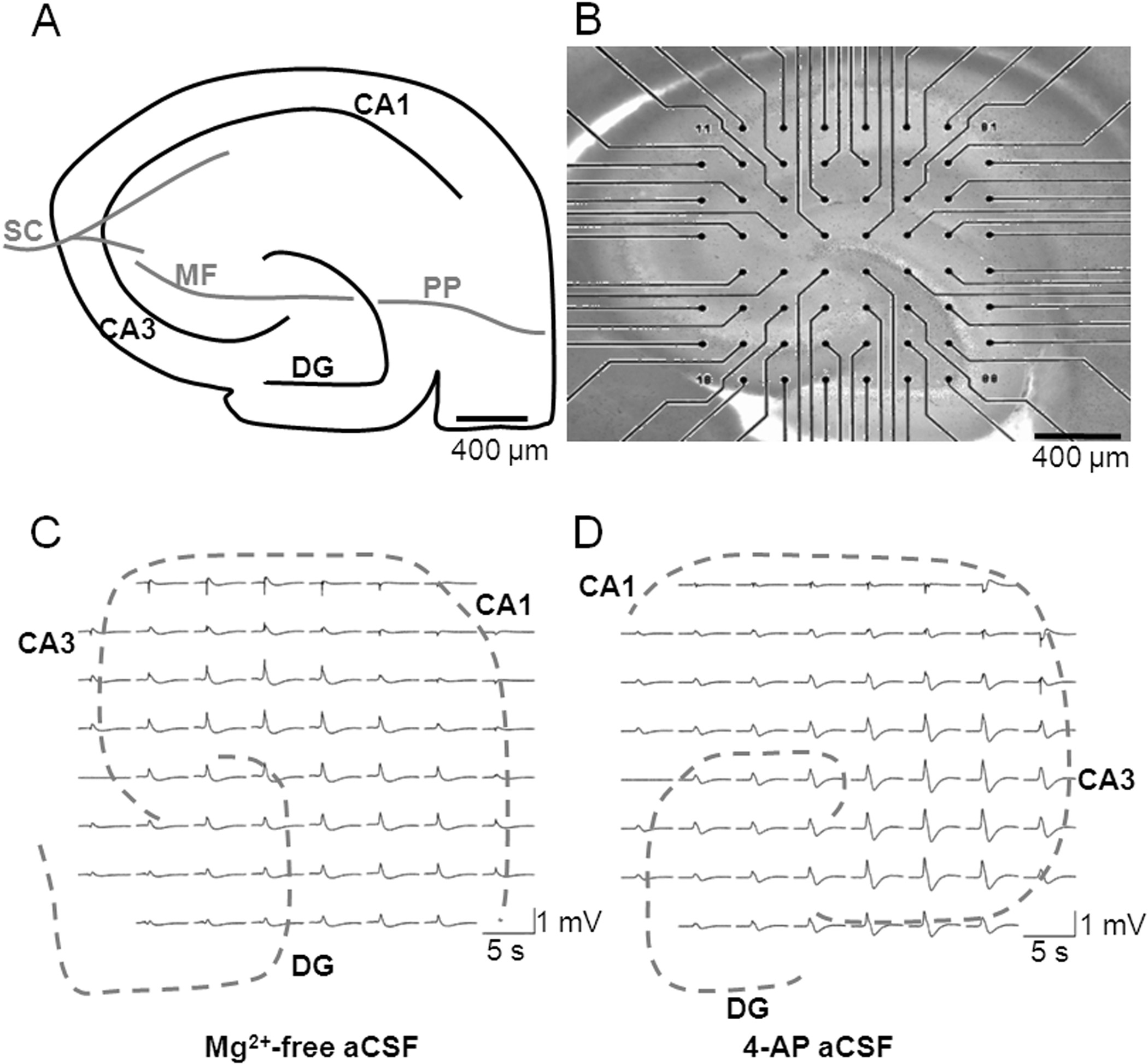
well defined architecture (Fig. 1A), exhibited no spontaneous
et al., 2008). All data are expressed as mean ⫾ S.E.M.
LFP events in control aCSF, and proved readily amenable to
Pharmacology. The following agents were used: AM251,
MEA recording (Fig. 1B). We sought to take advantage of the
WIN55,212-2 (Tocris-Cookson, Bristol, UK), CBD (GW Pharmaceu-
ability of MEAs to record spatiotemporal activity at multiple
ticals), and [3H]SR141716A and [35S]GTP␥S (GE Healthcare, Chal-
discrete, identifiable regions by investigating activity at CA1,
font St. Giles, Buckinghamshire, UK). All other reagents were from
CA3, and DG regions within the hippocampus. Application of
Mg2⫹-free aCSF (Fig. 1C) or 4-AP aCSF (Fig. 1D) to hip-pocampal slices resulted in the appearance of robust sponta-
neous epileptiform LFPs across the preparation. LFPs were
Characterization of Mg2ⴙ-Free and 4-AP Models of
consistent with status epilepticus-like activity and were re-
Epileptiform Activity Using MEA Electrophysiology.
liably recorded using the multisite MEA technique (Table 1).
To investigate neuronal excitability in vitro, we used both the
Slice-to-slice variability and electrode contact variability re-
Mg2⫹-free and 4-AP models of epileptiform activity in acute
sulted in substantial variation in signal strength (Table 1);
hippocampal brain slices, as measured using MEA electro-
therefore, subsequent drug-induced changes in burst charac-
physiology. Two separate models of epileptiform activity
teristics were normalized to control bursts before drug appli-
were used to provide a broader analysis of drug effects (Hill
cation (these analyses are fully characterized in Hill et al.,
et al., 2009; Whalley et al., 2009). Hippocampal slices have a
Effects of CBD in the Mg2ⴙ-Free Model of in Vitro
Epileptiform Activity. We first examined the effects of
CBD in the Mg2⫹-free model to assess CBD effects on a
receptor-dependent model of epileptiform activity. The Mg2⫹-
free model removes the Mg2⫹-dependent block of NMDA
glutamate receptors, rendering them more responsive to syn-
aptically released glutamate at resting membrane potentials.
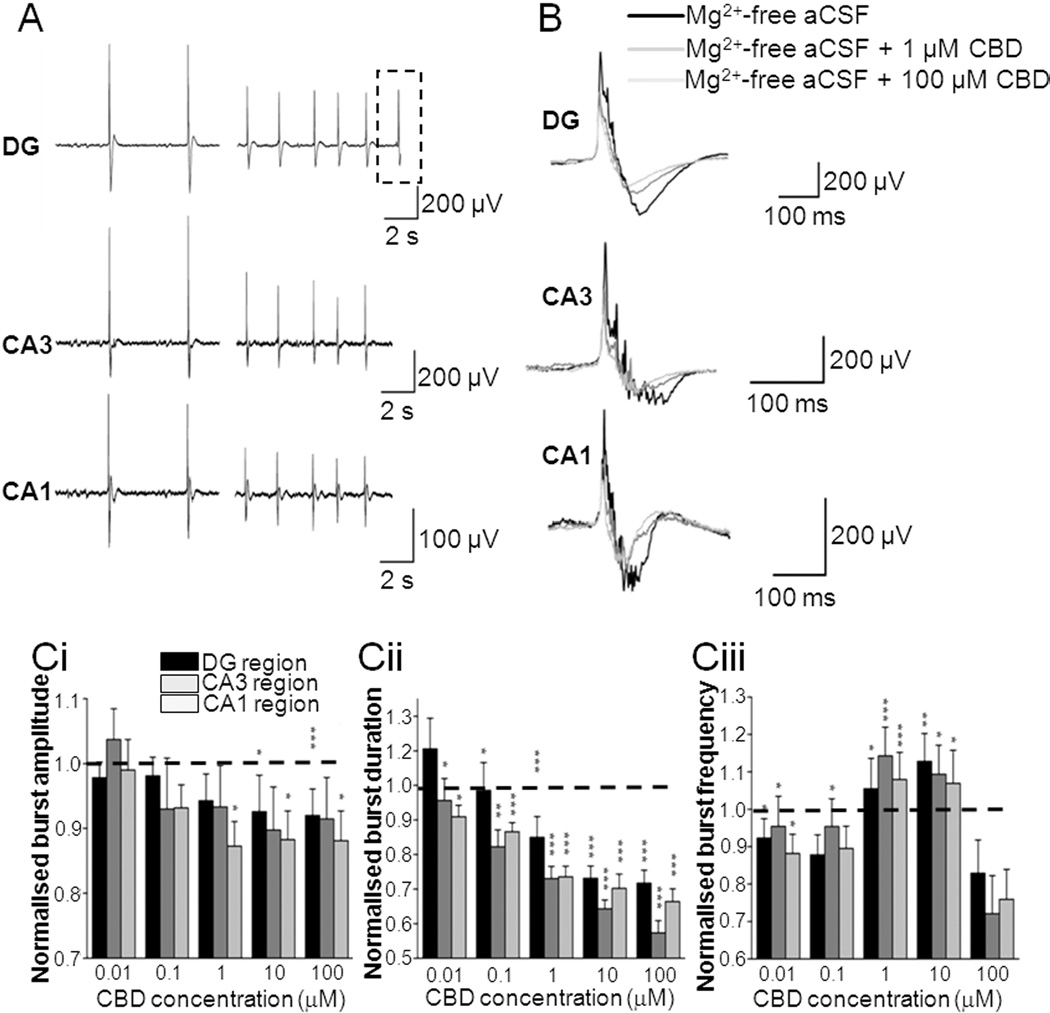
In Mg2⫹-free aCSF, CBD significantly decreased LFP burst
amplitude in the CA1 (1–100 M CBD) and DG (10 –100 M
CBD) regions (Fig. 2, A, B, and Ci). In contrast, CBD (0.01–
100 M) effects on LFP burst amplitude in CA3 failed to
reach significance. CBD decreased burst duration in CA1
(0.01–100 M CBD), CA3 (0.01–100 M CBD), and DG (0.1–
100 M CBD) regions (Fig. 2, A, B, and Cii). CBD (0.01–10
M) also caused an increase in burst frequency in all regionstested (Fig. 2Ciii; however, this effect was lost at 100 MCBD. To correlate these data with information on LFP burstinitiation and spread across the hippocampal brain slice, weconstructed contour plots (Fig. 3A) and associated video an-imations (Supplemental Fig. 1). Such plots spatiotemporallyvisualize the "8 ⫻ 8" MEA configuration (Fig. 1, B and C) andthe individual LFP activity shown in raw data traces (Fig. 2,A and B). In these experiments, Mg2⫹-free aCSF-induced
Fig. 1. Hippocampal slices are amenable to MEA recording. A, schematic
representation of hippocampal slice showing the position of CA1, CA3,
bursts typically originated in the CA3 region of the hip-
and DG regions, together with major pathways: Schaffer collateral (SC),
pocampal slice preparation and propagated along the princi-
mossy fiber (MF), and perforant pathway (PP). B, micrograph showing a
pal cell layer toward CA1. LFP events induced by Mg2⫹-free
hippocampal brain slice (stained with pontamine blue) mounted onto asubstrate-integrated MEA (60 electrodes of 30 m diameter, spaced 200
aCSF had a mean propagation speed of 0.229 ⫾ 0.048 m/s
m apart in an ⬃8 ⫻ 8 arrangement). Scale bar, 400 m. Representative
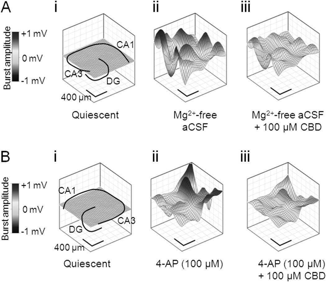
(n ⫽ 6). CBD (100 M) caused a clear suppression of Mg2⫹-
LFP burst activity was recorded at 60 electrodes across a hippocampal
free-induced LFP burst amplitude peak source and peak sink
slice in (C) Mg2⫹-free aCSF and (D) 4-AP aCSF. Traces were high pass-filtered in an MC_rack at 2 Hz.
values across the hippocampal slice (Fig. 3A; Supplemental
TABLE 1Characterization measures for the Mg2⫹-free and 4-AP (100 M) aCSF-induced LFP epileptiform activity in the CA1, CA3, and DG regions of thehippocampusLFP peak burst amplitude values are presented as a minimum and maximum range and mean ⫾ S.E.M. LFP burst duration and frequencies are presented as mean ⫾ S.E.M.
A minimum of six separate hippocampal slices were used in the characterisation of each in vitro model.
LFP Peak Burst Amplitude
Hippocampal Region
LFP Burst Duration
LFP Burst Frequency
Mean ⫾ S.E.M.
CA1 (n ⫽ 15)
CA3 (n ⫽ 12)
DG (n ⫽ 15)
CA1 (n ⫽ 15)
CA3 (n ⫽ 13)
DG (n ⫽ 18)
Cannabidiol as an Antiepileptic Agent
Fig. 1). Propagation speed across the brain slice in Mg2⫹-freeaCSF was not affected by 100 M CBD (0.232 ⫾ 0.076 m/s;n ⫽ 6; P ⬎ 0.5). Taken together, these data suggest that,although CBD attenuates epileptiform LFP amplitude andduration in the Mg2⫹-free model, the rate of signal spreadacross the preparation is not changed (see Discussion).
Effects of CBD in the 4-AP Model of in Vitro Epilep-
tiform Activity. We next examined the effects of CBD on
epileptiform bursting events in the 4-AP model of status
epilepticus-like activity. 4-AP acts to block postsynaptic volt-
age-dependent K⫹ channels and inhibits neuronal repolar-
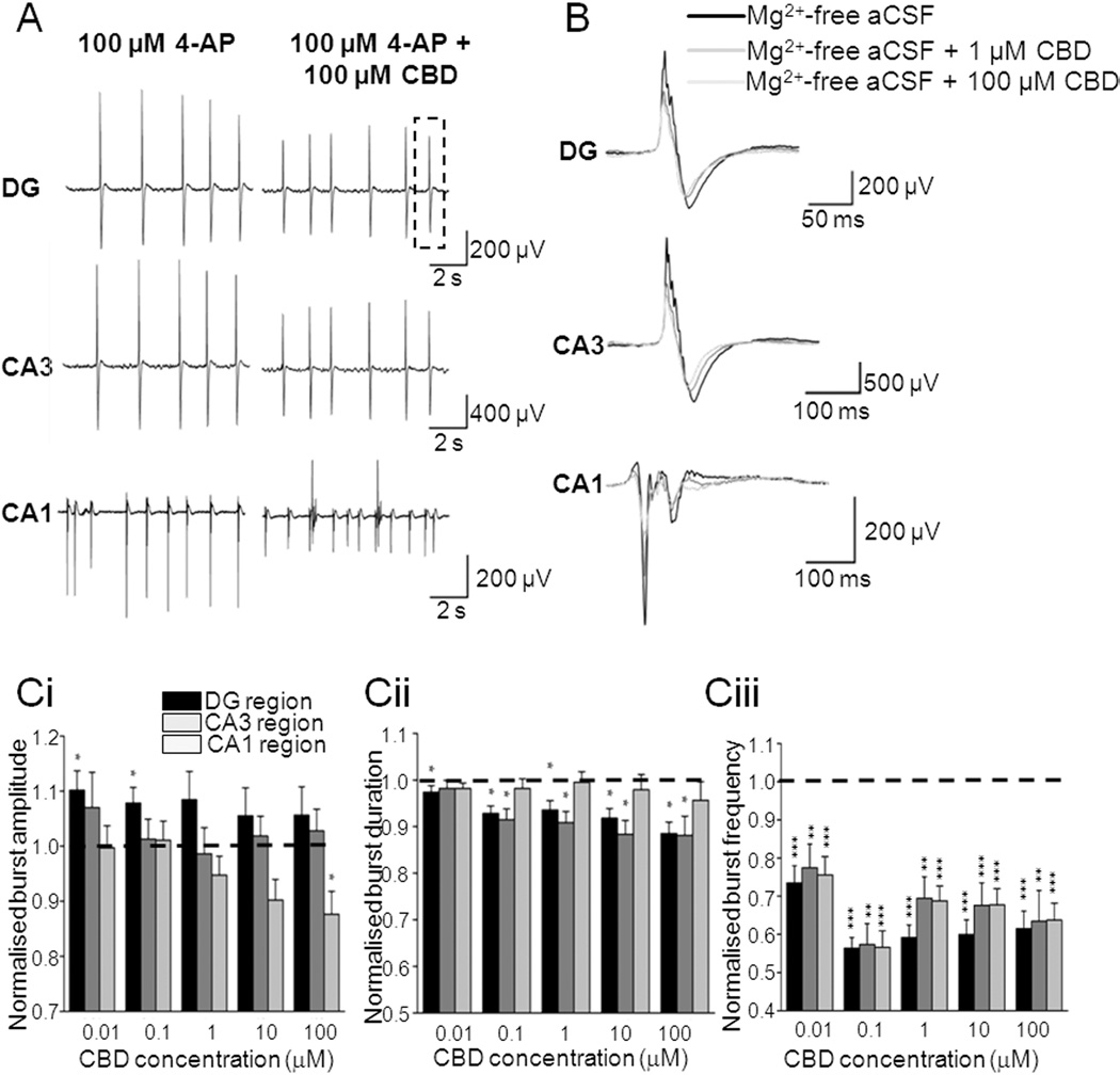
ization to effectively increase excitability. In 100 M 4-AP
aCSF, CBD (100 M) caused a significant decrease in LFP
burst amplitude in CA1 only (Fig. 4, A, B, and Ci). In con-
trast, CBD (0.01– 0.1 M) caused an unexpected small, but
significant, increase in LFP burst amplitude in the DG,
which was not apparent at higher CBD concentrations in this
or other hippocampal regions (Fig. 4Ci). CBD caused a de-
crease in burst duration in the DG (0.01–100 M CBD) and
CA3 (0.1–100 M CBD) but was without an overall effect on
CA1 (Fig. 4, A, B, and Cii). CBD (0.01–100 M) also caused a
significant decrease in burst frequency in all regions tested
(Fig. 4Ciii). In the same manner as for the Mg2⫹-free model,
Fig. 2. CBD attenuates epileptiform activity induced by Mg2⫹-free aCSF.
contour plots of 4-AP-induced epileptiform LFP burst events
A, representative traces showing the effects of 100 M CBD on Mg2⫹-free
(Fig. 3B) permitted spatiotemporal visualization of activity
aCSF-induced LFP bursts in different regions of hippocampal slices.
Dotted lines represent an individual LFP (as shown in B). B, effects of 1
across the slice preparation (Supplemental Fig. 1). 4-AP
and 100 M CBD on a representative individual Mg2⫹-free aCSF-induced
aCSF-induced bursts typically were initiated in CA3 before
LFP burst. C, bar graphs showing the effects of acute treatment of
spreading to CA1 with a propagation speed of 0.146 ⫾ 0.033
increasing CBD concentrations on normalized burst amplitude (Ci), nor-malized burst duration (Cii), and normalized burst frequency in Mg2⫹-free aCSF (Ciii). Note that burst amplitudes have been adjusted forrun-down and burst frequencies have been adjusted for run-up as de-scribed under Materials and Methods. Values are means ⫾ S.E.M. for thelast 10 LFP bursts in each condition. ⴱ, P ⱕ 0.05; ⴱⴱ, P ⱕ 0.01; ⴱⴱⴱ, P ⱕ0.001 (two-tailed Mann-Whitney U test).
Fig. 4. CBD attenuates epileptiform activity induced by 4-AP aCSF. A,
representative traces showing effects of 100 M CBD on 4-AP aCSF-
induced LFP bursts in different regions of a hippocampal slice. Dotted
Fig. 3. CBD attenuates epileptiform activity induced by Mg2⫹-free and
lines represent an individual LFP (as shown in B). B, effects of 1 and 100
4-AP aCSF. Representative contour plots illustrating CBD effects upon
M CBD on a representative individual 4-AP aCSF-induced LFP burst.
spatiotemporal epileptiform burst features. A, in the continued presence
C, bar graphs showing the effects of acute treatment of increasing CBD
of Mg2⫹-free aCSF: quiescent period between epileptiform burst events
concentrations on normalized burst amplitude (Ci), normalized burst
also showing hippocampal slice orientation (i), peak source in the absence
duration (Cii), and normalized burst frequency in the 4-AP aCSF (Ciii).
of CBD (ii), and peak source in the presence of CBD (100 M) (iii). B, in
Note that burst amplitudes have been adjusted for run-down and burst
the continued presence of 100 M 4-AP: quiescent period between epi-
frequencies have been adjusted for run-up as described under Materials
leptiform burst events also showing hippocampal slice orientation (i),
and Methods. Values are means ⫾ S.E.M. for the last 10 LFP bursts in
peak source in the absence of CBD (ii), and peak source in the presence of
each condition. ⴱ, P ⱕ 0.05; ⴱⴱ, P ⱕ 0.01; ⴱⴱⴱ, P ⱕ 0.001 (two-tailed
CBD (100 M) (iii).
Mann-Whitney U test).
Jones et al.
m/s (n ⫽ 5). CBD (100 M) caused a clear suppression of
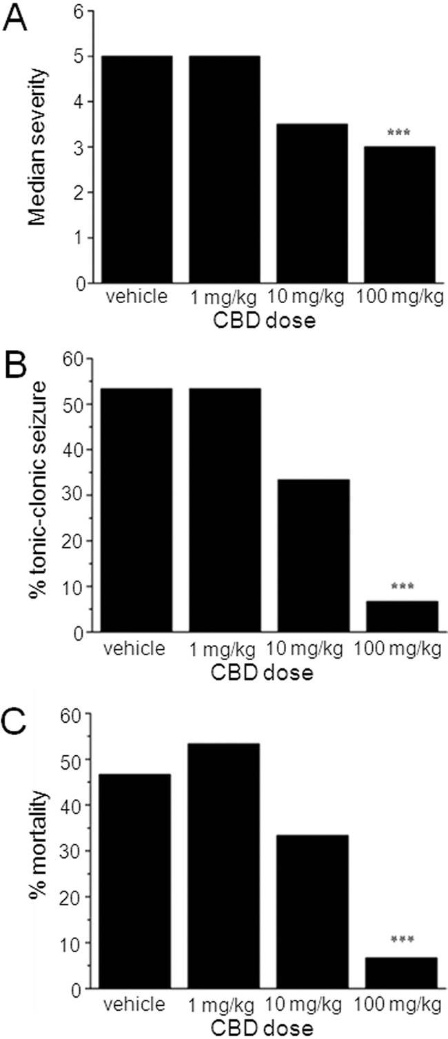
seizure states were observed. In contrast to the lack of defin-
4-AP-induced epileptiform LFP burst amplitude (Fig. 3B;
itive effects on seizure latency, CBD (100 mg/kg) demon-
Supplemental Fig. 1). Propagation speed across the brain
strated clear anticonvulsant effects via measures of seizure
slice in 4-AP aCSF was not affected by 100 M CBD (0.176 ⫾
severity (Fig. 6, A and B) and mortality (Fig. 6C). When the
0.046 m/s, n ⫽ 6; P ⬎ 0.5).
severity of PTZ-induced seizures is considered, vehicle-
Taken together, these data show that CBD displayed clear
treated animals reached a median score of 5 (tonic-clonic
concentration-related, region-specific, anticonvulsant prop-
seizures with a loss of righting reflex), the most severe on the
erties in two different in vitro models of epileptiform activity,
scoring scale (Fig. 6A). In contrast, animals treated with 100
attenuating LFP burst amplitude and duration, but with no
mg/kg CBD exhibited a significantly reduced median score of
effect on the rate of signal propagation in either model.
3.5 (forelimb clonus with a tonic component, but with the
Effects of CBD in the PTZ Model of Generalized Sei-
righting reflex preserved; n ⫽ 15 animals; P ⬍ 0.001) (Fig.
zures. We next assessed the effects of CBD (1, 10, and 100
6A). This was associated with a marked decrease in the
mg/kg i.p.) on PTZ-induced generalized seizures in adult
proportion of animals that developed the most severe tonic-
male rats. PTZ acts as a GABA receptor antagonist and this
clonic seizures, which was reduced from 53% in vehicle to 7%
model is well defined and used as a standard for the identi-
by 100 mg/kg CBD (n ⫽ 15 animals, P ⬍ 0.001) (Fig. 6B).
fication of potential anticonvulsants to treat generalized
Finally, percent mortality was significantly reduced from
clonic seizures (Lo¨scher et al., 1991). Seizures were defined
47% in vehicle to 7% by 100 mg/kg CBD (n ⫽ 15 animals, P ⬍
by a standard scoring scale (see Materials and Methods).
0.001) (Fig. 6C). Overall, these in vivo data confirm our in
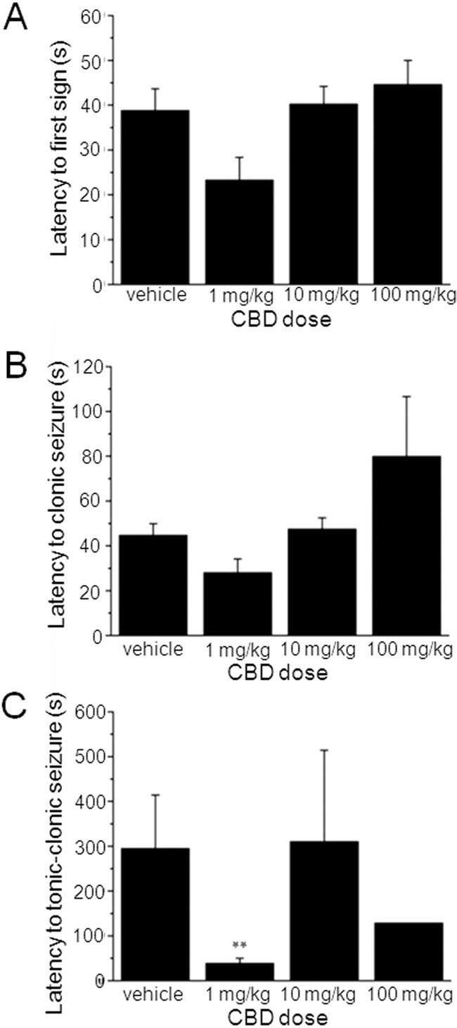
CBD at any dose did not significantly alter the latency to the
vitro results above and fully support an anticonvulsant ac-
first sign of PTZ-induced seizures (Fig. 5A) or latency to
tion for CBD.
development of clonic (Fig. 5B) seizures. Unexpectedly, CBD
Effects of CBD in Receptor Binding Assays. It is
(1 mg/kg) reduced latency to tonic-clonic seizures (P ⬍ 0.01)
known that hippocampal CB receptor expression on gluta-
(Fig. 5C). No other effects of CBD on latency to specific
matergic terminals is selectively down-regulated under epi-
Fig. 5. CBD has no clear effects on seizure latency in vivo. Bar graphs
showing lack of effects of CBD (1, 10, and 100 mg/kg) on latency to the
Fig. 6. CBD reduces seizure severity and mortality in vivo. Bar graphs
first sign of a seizure (A), latency to clonic seizures (B), and latency to
showing effects of CBD (1, 10, and 100 mg/kg) on median seizure severity
tonic-clonic seizures (C). Each data set n ⫽ 15 animals. Note that CBD (1
(A), percentage of animals reaching tonic-clonic seizures (B), and percent
mg/kg) reduced latency to tonic-clonic seizures; this proconvulsant action
mortality (C). Each data set n ⫽ 15 animals. CBD (100 mg/kg) signifi-
was not observed at higher CBD doses. ⴱⴱ, P ⱕ 0.01 (one-way analysis of
cantly reduced all of these parameters: ⴱⴱⴱ, P ⱕ 0.001 (nonparametric
variance) with a post hoc Tukey test).
binomial test).
Cannabidiol as an Antiepileptic Agent
leptic conditions (Luda´nyi et al., 2008); moreover, activation
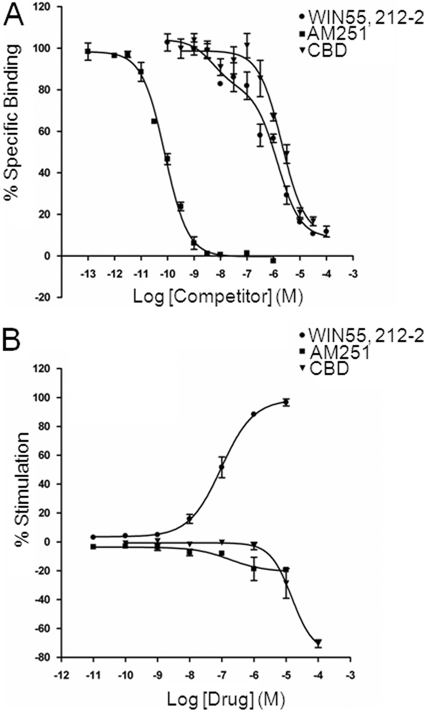
Finally, we investigated potential functional effects of CBD
receptors by eCBs is protective against seizures
using [35S]GTP␥S binding assays in rat cortical membranes;
(Monory et al., 2006) and exogenous CB agonists decrease
CBD actions were compared with effects of WIN55,212-2 and
epileptiform activity in hippocampal neurons (Shen and
AM251 (Fig. 7B). We first confirmed the presence of functional
Thayer, 1999; Blair et al., 2006). Therefore, we determined
CB receptors. Accordingly, WIN55,212-2 caused an increase in
potential CBD actions at CB receptors. Competition binding
percent stimulation of [35S]GTP␥S binding with an EC
assays were performed for CBD against the CB receptor
95.1 ⫾ 0.1 nM (n ⫽ 3); for 10 M WIN55,212-2, E
antagonist [3H]SR141716A in isolated cortical membranes;
3.5% (n ⫽ 3). AM251 had no stimulatory effect on
CBD effects were compared with those of the standard syn-
[35S]GTP␥S binding at tested concentrations of ⬍1 M; at
thetic CB receptor agonist WIN55,212-2 and the CB recep-
micromolar concentrations AM251 caused a moderate de-
tor antagonist AM251 (Fig. 7A). AM251 displacement of
pression of [35S]GTP␥S binding (10 M AM251: ⫺20.3 ⫾
[3H]SR141716A binding occurred with high affinity (K ⫽
4.3%, n ⫽ 3). CBD had no effect at concentrations ⱕ1 M;
190 ⫾ 56 pM; n ⫽ 4) and was best fitted by a one-site
large decreases in [35S]GTP␥S binding were seen at 10 M
competition model (Hill slope ⫽ ⫺1.08 ⫾ 0.13; n ⫽ 4). In
CBD (⫺28.8 ⫾ 10.3%, n ⫽ 3) and 100 M CBD (⫺76.7 ⫾
contrast, WIN55,212-2 displacement was best fitted to a two-
15.9%, n ⫽ 3).
site model with a high-affinity site (K ⫽ 7.03 ⫾ 4.1 nM; %
Thus, overall, CBD had clear antiepileptogenic and anti-
R ⫽ 27.4 ⫾ 5.0%; n ⫽ 4) and a low-affinity site (K ⫽ 904 ⫾
seizure effects but only low affinity and no clear agonist
155 nM; n ⫽ 4); in these experiments, Hill slopes for either
effects at cortical CB receptors.
the low- or high-affinity site did not match unity. CBD dis-placement of [3H]SR141716A occurred with low affinity (K ⫽
1.82 ⫾ 0.38 M; n ⫽ 4) and was best fitted by a one-site model(Hill slope ⫽ ⫺1.15 ⫾ 0.11; n ⫽ 4).
CBD Reduces Excitability in in Vitro Models of Epi-
leptiform Activity. In the present study, we use extracel-
lular MEA recordings to demonstrate that CBD attenuates
epileptiform activity in both the Mg2⫹-free and 4-AP in vitro
models of status epilepticus in the mammalian hippocampus,
a prominent epileptogenic region (Ben-Ari and Cossart,
2000). The major effects of CBD were to decrease LFP burst
amplitude and duration in a hippocampal region-specific
manner. In general, the CA1 region was most sensitive to
CBD effects. Thus, LFP amplitude was significantly reduced
at lower CBD concentrations in CA1 than in CA3 (with DG
remaining unaffected) in Mg2⫹-free aCSF, and CA1 was the
only region in which LFP amplitude was affected in 4-AP aCSF.
This is of interest because the CA1 region represents the major
output of the hippocampus, relaying information to cortical and
subcortical sites and is intimately involved in propagation of
epileptic activity (McCormick and Contreras, 2001). Contour
plots constructed from data in the Mg2⫹-free and 4-AP models
confirmed that LFP bursts typically originated in the CA3 re-
gion and propagated toward CA1 (Feng and Durand, 2005),
strongly suggesting that CA1 is a major focus of epileptic activ-
ity in the two models used and illustrating that CBD exerts a
significant antiepileptiform effect in this region.
Overall, CBD induced more prominent effects in Mg2⫹-free
than in 4-AP aCSF. This result may reflect inherent differencesbetween the two models, which affect NMDA glutamate recep-tors and K⫹ channels, respectively. CBD had contrasting ac-tions on LFP burst frequency between models; frequency wasincreased in all regions by CBD in Mg2⫹-free aCSF but wasdecreased in all regions in 4-AP aCSF. It is interesting to notethat 100 M CBD was without effect on burst frequency in theMg2⫹-free model, in contrast to data for all the lower concen-trations of CBD tested. This finding was the only indication of
Fig. 7. CBD displaces [3H]SR141716A binding with low affinity and
any biphasic action of CBD, a common phenomenon associated
lacks agonist effects in [35S]GTP␥S binding assays in cortical mem-
with cannabinoids whereby increasing concentrations cause
branes. A, representative competition curves for the CB receptor agonist
changes in the pharmacological "direction" of action (Pertwee,
WIN55,212-2, the selective CB receptor antagonist AM251, and CBD
against 1 nM [3H]SR141716A (a selective CB receptor antagonist) bind-
ing to cortical membranes. Points are means ⫾ S.E.M. of triplicate points.
In light of the region-specific effects of CBD, it will be of
B, agonist-binding curves for the CB receptor agonist WIN55,212-2, the
interest in the future to investigate the cellular mechanisms
receptor antagonist AM251, and CBD stimulation of
of action of CBD using intracellular recording from individ-
[35S]GTP␥S binding to cortical membranes. Points are means ⫾ S.E.M. oftriplicate points from three separate experiments.
ual neurons in selected hippocampal regions. In both the
Jones et al.
Mg2⫹-free and 4-AP models, contour plots and subsequent
that CBD has no stimulatory agonist activity but that CBD at
analyses showed that CBD caused clear attenuation in LFP
micromolar concentrations decreases G protein activity. These
burst amplitude but had no overall effect on burst propaga-
findings are also in agreement with studies in mouse whole
tion speed. These findings suggest that CBD acts to reduce
brain membranes (Thomas et al., 2007), which showed that
the magnitude of epileptiform activity while leaving speed of
CBD has only low affinity at CB and CB receptors but acts
information transmission across the hippocampal slice in-
efficaciously as an antagonist at both receptor types (Thomas et
tact. It is possible that this action may result in a more
al., 2007; see Pertwee, 2008). There are a number of potential
tolerable side effect profile for CBD in comparison with ex-
mechanisms by which ligands acting at CB receptors may me-
isting AEDs if used in a clinical setting.
diate anticonvulsant effects. Receptor agonists may act at CB1
CBD Has Anticonvulsant Properties in the PTZ
on excitatory presynaptic terminals to inhibit glutamate neuro-
Model of Generalized Seizures. CBD had beneficial ef-
transmitter release. Such a mechanism is unlikely here as CBD
fects on seizure severity and lethality in response to PTZ
has no agonist effect in GTP␥S binding assays (Thomas et al.,
administration without delaying the time taken for seizures
2007). An alternative is that antagonists act at CB receptors on
to develop. CBD (100 mg/kg) demonstrated clear anticonvul-
inhibitory presynaptic terminals to increase GABA release. We
sant effects in terms of significant reductions in median
have demonstrated such a mechanism for the phytocannabi-
seizure severity, tonic-clonic seizures, and mortality. Partic-
noid ⌬9-tetrahydrocannabivarin in the cerebellum (Ma et al.,
ularly striking effects were that ⬍10% of animals developed
2008), where displacement of eCB tone may lead to increased
tonic-clonic seizures or died when treated with CBD in com-
inhibition. It may also be speculated that the decreases in G
parison to approximately 50% of vehicle-treated animals.
protein activity seen in GTP␥S binding assays represent an
The present data strongly substantiate a number of earlier in
inverse agonist action at CB receptors; for example, if CBD
vivo studies suggesting that CBD has anticonvulsant poten-
were acting as an inverse agonist at CB receptors on inhibitory
tial (Lutz, 2004; Scuderi et al., 2009). CBD has been reported
presynaptic terminals an increase in GABA release could lead
to have relatively potent anticonvulsant action in maximal
to reduced excitability. However, mechanisms involving in-
electroshock (a model of partial seizure with secondary gen-
creases in GABA release are unlikely here as CBD was effective
eralization) (Karler et al., 1974; Consroe and Wolkin, 1977).
in reducing seizures in vivo in the presence of the GABAA
Moreover, CBD prevented tonic-clonic seizures in response to
receptor antagonist PTZ. Moreover, CBD-induced reductions in
electroshock current (Consroe et al., 1982). There are limited
[35S]GTP␥S binding to whole brain membranes were retained
clinical data on CBD effects on seizure frequency in humans
in CB knockout [cnr1(⫺/⫺)] mice, suggesting that CBD is not
(Gordon and Devinsky, 2001). However, in one small double-
an inverse agonist at CB receptors (Thomas et al., 2007).
blind study of eight patients with uncontrolled secondary
Overall, the low affinity and lack of agonist activity at CB1
generalized epilepsy treated with 200 to 300 mg of CBD, four
receptors suggests that the CBD anticonvulsant effects re-
remained symptom-free and three had signs of improvement
ported here are potentially mediated by CB receptor-indepen-
(Cunha et al., 1980). One potential concern was the high
dent mechanisms. In addition to the study of Thomas et al.,
doses of CBD used by Cunha et al. (1980). Because all new
showing that CBD actions were unaltered in cnr1(⫺/⫺) mice,
therapies must be introduced initially in an adjunct capacity
CBD anticonvulsant effects in the maximal electroshock model
to existing medication, the present study suggests that one
were unaffected by the CB receptor antagonist SR141716A,
attractive possibility is a role for CBD as an adjunct in
whereas those of ⌬9-THC and WIN55,212-2 were blocked
generalized seizures. In this regard, earlier animal studies
(Wallace et al., 2001).
indicate that CBD enhances the effects of phenytoin (al-
In addition to CB receptors, a number of alternative molecu-
though CBD reduced the potency of other AEDs) (Consroe
lar targets may also contribute to CBD effects on neuronal
and Wolkin, 1977). In the future, it will be of interest to
excitability. CBD has been reported to be an antagonist at
extend studies to other animal seizure models and also to
GPR55, a non-CB /CB receptor (Ryberg et al., 2007); in con-
combination therapies with selective AEDs to determine the
trast, a recent study demonstrated that CBD has no effect at
full clinical anticonvulsant potential of CBD against a range
GPR55 (Kapur et al., 2009). CBD may cause an increase in
of epilepsy phenotypes.
anticonvulsant eCBs via the reported inhibition of the catabolic
Mechanism of Action. Cannabinoid actions are mediated
enzyme fatty acid hydrolyase, which degrades anandamide,
by CB and CB receptors, potentially by the GPR55 receptor,
and/or the blockade of anandamide uptake (Watanabe et al.,
and also by cannabinoid receptor-independent mechanisms
1996; Rakhshan et al., 2000; Bisogno et al., 2001). CBD is
(Howlett et al., 2004; Ryberg et al., 2007). In regard to epilepsy,
reported to be a weak agonist at human TRPV1 receptors
CB receptors are densely expressed in the hippocampal forma-
(Bisogno et al., 2001); a more recent study suggests an action for
tion (Herkenham et al., 1990; Tsou et al., 1998) where their
CBD at rat and human transient receptor potential vanilloid 2
activation is widely reported to be antiepileptic in animal mod-
but not rat transient receptor potential vanilloid 1 receptors
els (Shen and Thayer, 1999; Wallace et al., 2001; but see Clem-
(Qin et al., 2008). More relevant to potential effects on neuronal
ent et al., 2003). Here, we demonstrate that CBD displaced the
excitability in the CNS is the demonstration that CBD exerts a
selective CB receptor antagonist [3H]SR141716A in cortical
bidirectional action on [Ca2⫹] levels in hippocampal neurons
membranes with relatively low affinity (K ⫽ 1.82 M); these
(Ryan et al., 2009). Under control conditions, CBD induces
data are in line with values reported in whole brain membranes
increases in [Ca2⫹] ; in contrast, in the presence of 4-AP (which
(reviewed in Pertwee, 2008) and our data in cerebellar mem-
induces seizure-like [Ca2⫹] oscillations) or increased extracel-
branes (Smith et al., 2009). CB receptor/G-protein coupling may
lular K⫹, CBD acts to reduce [Ca2⫹] and thus epileptiform
differ among distinct brain regions (Breivogel et al., 1997; Den-
activity, via an action on mitochondria Ca2⫹ stores. A further
nis et al., 2008); therefore, we investigated CBD effects on
recent report provides the first evidence that CBD can also
[35S]GTP␥S binding in isolated cortical membranes. We showed
block low-voltage-activated (T-type) Ca2⫹ channels (Ross et al.,
Cannabidiol as an Antiepileptic Agent
2008), important modulators of neuronal excitability. Finally,
Atypical responsiveness of the orphan receptor GPR55 to cannabinoid ligands.
J Biol Chem 284:29817–29827.
CBD may also enhance the activity of inhibitory glycine
Karler R, Cely W, and Turkanis SA (1974) Anticonvulsant properties of ⌬9-
receptors (Ahrens et al., 2009). Overall, the demonstration
tetrahydrocannabinol and other cannabinoids. Life Sci 15:931–947.
that CBD acts on multiple molecular targets that each play a
Kwan P and Brodie MJ (2007) Emerging drugs for epilepsy. Expert Opin Emerg
Drugs 12:407– 422.
key role in neuronal excitability reinforces the potential of
Lo¨scher W, Ho¨nack D, Fassbender CP, and Nolting B (1991) The role of technical,
CBD as an AED.
biological and pharmacological factors in the laboratory evaluation of anticonvul-
sant drugs. III. Pentylenetetrazole seizure models. Epilepsy Res 8:171–189.
In conclusion, our data in separate in vitro models of epilepti-
Lowry OH, Rosebrough NJ, Farr AL, and Randall RJ (1951) Protein measurement
form activity and, in particular, the beneficial reductions in seizure
with the Folin phenol reagent. J Biol Chem 193:265–275.
Luda´nyi A, Eross L, Czirja´k S, Vajda J, Hala´sz P, Watanabe M, Palkovits M,
severity caused by CBD in an in vivo animal model of generalized
Maglo´czky Z, Freund TF, and Katona I (2008) Downregulation of the CB1 canna-
seizures suggests that earlier, small-scale clinical trials for CBD in
binoid receptor and related molecular elements of the endocannabinoid system in
epileptic human hippocampus. J Neurosci 28:2976 –2990.
untreated epilepsy warrant urgent renewed investigation.
Lutz B (2004) On-demand activation of the endocannabinoid system in the control of
neuronal excitability and epileptiform seizures. Biochem Pharmacol 68:1691–1698.
Ma YL, Weston SE, Whalley BJ, and Stephens GJ (2008) The phytocannabinoid
⌬9-tetrahydrocannabivarin modulates inhibitory neurotransmission in the cere-
We thank Professor Philip Strange for useful discussion and Colin
bellum. Br J Pharmacol 154:204 –215.
Stott (GW Pharmaceuticals) and Professor Gernot Riedel (University
McCormick DA and Contreras D (2001) On the cellular and network bases of
epileptic seizures. Annu Rev Physiol 63:815– 846.
of Aberdeen) for pharmacokinetics data.
Mechoulam R and Shvo Y (1963) Hashish. I. The structure of cannabidiol. Tetrahe-
Monory K, Massa F, Egertova´ M, Eder M, Blaudzun H, Westenbroek R, Kelsch W,
Abramoff MD, Magelhaes PJ, and Ram SJ (2004) Image processing with ImageJ.
Jacob W, Marsch R, Ekker M, et al. (2006) The endocannabinoid system controls
J Biophoton Int 11:36 – 42.
key epileptogenic circuits in the hippocampus. Neuron 51:455– 466.
Ahrens J, Demir R, Leuwer M, de la Roche J, Krampfl K, Foadi N, Karst M, and
Pertwee RG (2008) The diverse CB1 and CB2 receptor pharmacology of three plant
Haeseler G (2009) The nonpsychotropic cannabinoid cannabidiol modulates and
cannabinoids: ⌬9-tetrahydrocannabinol, cannabidiol and ⌬9-tetrahydrocannabiva-
directly activates ␣1 and ␣1 glycine receptor function. Pharmacology 83:217–222.
rin. Br J Pharmacol 153:199 –215.
Ben-Ari Y and Cossart R (2000) Kainate, a double agent that generates seizures: two
Pohl M and Mares P (1987) Effects of flunarizine on metrazol-induced seizures in
decades of progress. Trends Neurosci 23:580 –587.
developing rats. Epilepsy Res 1:302–305.
Bhattacharyya S, Fusar-Poli P, Borgwardt S, Martin-Santos R, Nosarti C, O'Carroll
Qin N, Neeper MP, Liu Y, Hutchinson TL, Lubin ML, and Flores CM (2008) TRPV2
C, Allen P, Seal ML, Fletcher PC, Crippa JA, et al. (2009) Modulation of medio-
is activated by cannabidiol and mediates CGRP release in cultured rat dorsal root
temporal and ventrostriatal function in humans by ⌬9-tetrahydrocannabinol: a
ganglion neurons. J Neurosci 28:6231– 6238.
neural basis for the effects of Cannabis sativa on learning and psychosis. Arch Gen
Rakhshan F, Day TA, Blakely RD, and Barker EL (2000) Carrier-mediated uptake of
Psychiatry 66:442– 451.
the endogenous cannabinoid anandamide in RBL-2H3 cells. J Pharmacol Exp Ther
Bisogno T, Hanus L, De Petrocellis L, Tchilibon S, Ponde DE, Brandi I, Moriello AS,
Davis JB, Mechoulam R, and Di Marzo V (2001) Molecular targets for cannabidiol
Ross HR, Napier I, and Connor M (2008) Inhibition of recombinant human T-type
and its synthetic analogues: effect on vanilloid VR1 receptors and on the cellular
calcium channels by ⌬9-tetrahydrocannabinol and cannabidiol. J Biol Chem 283:
uptake and enzymatic hydrolysis of anandamide. Br J Pharmacol 134:845– 852.
16124 –16134.
Blair RE, Deshpande LS, Sombati S, Falenski KW, Martin BR, and DeLorenzo RJ
Ryan D, Drysdale AJ, Lafourcade C, Pertwee RG, and Platt B (2009) Cannabidiol
(2006) Activation of the cannabinoid type-1 receptor mediates the anticonvulsant
targets mitochondria to regulate intracellular Ca2⫹ levels. J Neurosci 29:2053–2063.
properties of cannabinoids in the hippocampal neuronal culture models of acquired
Ryberg E, Larsson N, Sjo¨gren S, Hjorth S, Hermansson NO, Leonova J, Elebring T,
epilepsy and status epilepticus. J Pharmacol Exp Ther 317:1072–1078.
Nilsson K, Drmota T, and Greasley PJ (2007) The orphan receptor GPR55 is a
Breivogel CS, Sim LJ, and Childers SR (1997) Regional differences in cannabinoid
novel cannabinoid receptor. Br J Pharmacol 152:1092–1101.
receptor/G-protein coupling in rat brain. J Pharmacol Exp Ther 282:1632–1642.
Sagredo O, Ramos JA, Decio A, Mechoulam R, and Ferna´ndez-Ruiz J (2007) Can-
Clement AB, Hawkins EG, Lichtman AH, and Cravatt BF (2003) Increased seizure
nabidiol reduced the striatal atrophy caused 3-nitropropionic acid in vivo by
susceptibility and proconvulsant activity of anandamide in mice lacking fatty acid
mechanisms independent of the activation of cannabinoid, vanilloid TRPV1 and
amide hydrolase. J Neurosci 23:3916 –3923.
adenosine A2A receptors. Eur J Neurosci 26:843– 851.
Consroe P, Benedito MA, Leite JR, Carlini EA, and Mechoulam R (1982) Effects of
Scuderi C, Filippis DD, Iuvone T, Blasio A, Steardo A, and Esposito G (2009)
cannabidiol on behavioral seizures caused by convulsant drugs or current in mice.
Cannabidiol in medicine: a review of its therapeutic potential in CNS disorders.
Eur J Pharmacol 83:293–298.
Phytother Res 23:597– 602.
Consroe P and Wolkin A (1977) Cannabidiol-antiepileptic drug comparisons and
Shen M and Thayer SA (1999) ⌬9-Tetrahydrocannabinol acts as a partial agonist to
interactions in experimentally induced seizures in rats. J Pharmacol Exp Ther
modulate glutamatergic synaptic transmission between rat hippocampal neurons
in culture. Mol Pharmacol 55:8 –13.
Cunha JM, Carlini EA, Pereira AE, Ramos OL, Pimentel C, Gagliardi R, Sanvito WL,
Smith I, Bevan SA, Whalley BJ, and Stephens GJ (2009) Phytocannabinoid affinities
Lander N, and Mechoulam R (1980) Chronic administration of cannabidiol to
at CB1 receptors in the mouse cerebellum. Proceedings of the 19th Annual Meeting
healthy volunteers and epileptic patients. Pharmacology 21:175–185.
of the International Cannabinoid Research Society; 2009 July 8 –11; Pheasant Run,
Dennis I, Whalley BJ, and Stephens GJ (2008) Effects of ⌬9-tetrahydrocannabivarin
St. Charles, IL. P81, International Cannabinoid Research Society.
on [35S]GTP␥S binding in mouse brain cerebellum and piriform cortex membranes.
Stett A, Egert U, Guenther E, Hofmann F, Meyer T, Nisch W, and Haemmerle H
Br J Pharmacol 154:1349 –1358.
(2003) Biological application of microelectrode arrays in drug discovery and basic
Egert U, Heck D, and Aertsen A (2002a) Two-dimensional monitoring of spiking
research. Anal Bioanal Chem 377:486 – 495.
networks in acute brain slices. Exp Brain Res 142:268 –274.
Straiker A and Mackie K (2005) Depolarization-induced suppression of excitation in
Egert U, Knott T, Schwarz C, Nawrot M, Brandt A, Rotter S, and Diesmann M
murine autaptic hippocampal neurones. J Physiol 569:501–517.
(2002b) MEA-Tools: an open source toolbox for the analysis of multi-electrode data
Thomas A, Baillie G, Philips AM, Razdan RK, Ross RA, and Pertwee RG (2007)
with MATLAB. J Neurosci Methods 117:33– 42.
Cannabidiol displays unexpectedly high potency as an antagonist of CB1 and CB2
Farrimond JA, Hill AJ, Jones NA, Stephens GJ, Whalley BJ, and Williams CM
receptor agonists in vitro. Br J Pharmacol 150:917–926.
(2009) A cost-effective high-throughput digital system for observation and acqui-
Tsou K, Brown S, San
˜ a MC, Mackie K, and Walker JM (1998) Immunohis-
sition of animal behavioral data. Behav Res Methods 41:446 – 451.
tochemical distribution of cannabinoid CB1 receptors in the rat central nervous
Feng Z and Durand DM (2005) Decrease in synaptic transmission can reverse the
system. Neuroscience 83:393– 411.
propagation direction of epileptiform activity in hippocampus in vivo. J Neuro-
Vivo M, Lin H, and Strange PG (2006) Investigation of cooperativity in the binding
physiol 93:1158 –1164.
of ligands to the D2 dopamine receptor. Mol Pharmacol 69:226 –235.
Gordon E and Devinsky O (2001) Alcohol and marijuana: effects on epilepsy and use
Wallace MJ, Martin BR, and DeLorenzo RJ (2002) Evidence for a physiological role
by patients with epilepsy. Epilepsia 42:1266 –1272.
of endocannabinoids in the modulation of seizure threshold and severity. Eur
Hampson AJ, Grimaldi M, Axelrod J, and Wink D (1998) Cannabidiol and (⫺)⌬9-
J Pharmacol 452:295–301.
tetrahydrocannabinol are neuroprotective antioxidants. Proc Natl Acad Sci U S A
Wallace MJ, Wiley JL, Martin BR, and DeLorenzo RJ (2001) Assessment of the
95:8268 – 8273.
role of CB1 receptors in cannabinoid anticonvulsant effects. Eur J Pharmacol
Herkenham M, Lynn AB, Little MD, Johnson MR, Melvin LS, de Costa BR, and Rice
KC (1990) Cannabinoid receptor localization in brain. Proc Natl Acad Sci U S A
Watanabe K, Kayano Y, Matsunaga T, Yamamoto I, and Yoshimura H (1996)
Inhibition of anandamide amidase activity in mouse brain microsomes by canna-
Hill AJ, Jones NA, Williams CM, Stephens GJ, and Whalley BJ (2009) Development
binoids. Biol Pharm Bull 19:1109 –1111.
of multi-electrode array screening for anticonvulsants in acute rat brain slices.
Whalley BJ, Stephens GJ, and Constanti A (2009) Investigation of the effects of the
J Neurosci Methods doi:10.1016/j.jneumeth.2009.10.007.
novel anticonvulsant compound carisbamate (RWJ-333369) on rat piriform corti-
Howlett AC, Breivogel CS, Childers SR, Deadwyler SA, Hampson RE, and Porrino
cal neurones in vitro. Br J Pharmacol 156:994 –1008.
LJ (2004) Cannabinoid physiology and pharmacology: 30 years of progress. Neu-
ropharmacology 47:345–358.
Address correspondence to: Dr. Gary Stephens, School of Pharmacy, Univer-
Iuvone T, Esposito G, De Filippis D, Scuderi C, and Steardo L (2009) Cannabidiol: a
sity of Reading, Whiteknights, P.O. Box 228, Reading RG6 6AJ, UK. E-mail:
promising drug for neurodegenerative disorders? CNS Neurosci Ther 15:65–75.
Kapur A, Zhao P, Sharir H, Bai Y, Caron MG, Barak LS, and Abood ME. (2009)
Source: http://www.v-cbc.ca/uploads/Cannabidiol%20displays%20antiepileptiform%20and%20antiseizure%20properties%20in%20vitro%20and%20in%20vivo.pdf
General information: Chronic Fatigue Syndrome Recognizing NASA's rocking results, it was later determined that rocking was also the best solution for CFS patients. Rocking provides easy, non-weight bearing, rhythmic motion and does a very important job in autonomic tone for a population that has difficulty accomplishing exercise. (Chronic Fatigue Syndrome affects approximately half a mil ion people in the US.)
Bioprocess Modelling for Learning Model Predictive Control (L-MPC) Mar´ıa Antonieta Alvarez1,�, Stuart M. Stocks2, and S. Bay Jørgensen1,�� 1 CAPEC, Department of Chemical and Biochemical Engineering, Technical University of Denmark, Søltofts Plads, 2800 Kgs. Lyngby, Denmark 2 Novozymes, Bagsværd, Denmark Abstract. Batch and Fed-Batch cultivation processes are used exten-sively in many industries where a major issue today is to reduce theproduction losses due to sensitivity to disturbances occurring betweenbatches and within batches. In order to ensure consistent product qualityby eliminating the influence of process disturbances it is very importantto consider implementation of monitoring and control and thereby signif-icantly improve the economic impact for these industries. A data drivenmodeling methodology is described for batch and fed batch processeswhich is based upon data obtained from operating processes. The chap-ter illustrates how additional production experiments may be designed toimprove model quality for control. The chapter also describes how the de-veloped models may be used for process monitoring, for ensuring processreproducibility through control and for optimizing process performanceby enforcing learning from previous batch runs through Learning ModelPredictive Control (L-MPC).