Kamagra gibt es auch als Kautabletten, die sich schneller auflösen als normale Pillen. Manche Patienten empfinden das als angenehmer. Wer sich informieren will, findet Hinweise unter kamagra kautabletten.
Ijcs.org
Five Fluorouracil, Hyaluronidase and Triamcinolone in the
Nasal Region.
Guillermo Blugermaniego Schavelzon, Gabriel Wexler
Department of Plastic and Reconstructive Surgery, Centros ByS, Buenos Aires,
Abstract: The use of five fluorouracil (5 FU) as antifibrotic started in the 1960s, in
hands of ophthalmologists, to prevent adherence after glaucoma and pterigion
surgery. In 1999 Fitzpatric presented his experience in keloids and hypertrophic scars,making a great contribution to their treatment. Fibroblasts main function is collagen
synthesis; in vicious scar the amount of collagen is normal, but what is altered is theratio between collagen subtypes. The use of triamcinolone, the previous standardtreatment produced different degrees of atrophy and telangiectasias. Infiltration isdone in the center of fibrosis, weekly for the first month, then every 15 days untilreaching the desired result. Softening, loss of volume and of retraction is seen sincethe first session. Also pain and itching disappears. Biopsy of treated scars hasorganized collagen fibers and less fibroblasts compared to non treated scars. Clinicalapplications: hypertrophic scars or keloids fixed to deep planes, "supratip" postrhinoplasty fibrosis, post rhinoplasty fibrosis (as preparation for other treatments),foreign body granulomas, and post burn retractions. The use in supratip deformitywhen secondary to rhinoplasty fibrotic scar has proven very effective, and also aspreparation for surgical (secondary rhinoplasty) and no surgical (bioplasty)procedures.Our experience in the treatment of nasal scars, fibrosis and retractionwith 5 FU is favorable. The results of infiltration with 5FU, hyaluronidase andtriamcinolone in low dose have clinical and histological demonstration of collagensynthesis reduction and reorganization of collagen cicatrizal fibers.
Keywords: corticosteroids, fibrosis, fibrosis histopathology, fibrosis treatment, five
fluorouracil, fibroblasts, foreign body granulomas, healing, hyaluronidase,
hypertrophic scar, keloids, nasal aesthetics, nasal dermatology, nasal deformity, nasal
reconstruction, post rhinplasty fibrosisburn sequelae, scar, scarring disorders, supratip
deformity.
Since Fitzpatrick[1] published his work with five fluorouracil (5FU) in 1999 weincluded it in our daily practice. The most frequent use of this drug is for treatment andprevention of hypertrophic scars, keloids and foreign body granulomas[2].
1
Address correspondence to Guillermo Blugerman: Department of Plastic and Reconstructive Surgery,
Centros ByS, Buenos Aires, Argentina; E-mail:
[email protected]
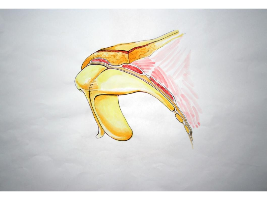
The particular anatomy of the skin in each nasal subunit makes scarring processcompletely different among them (Fig.1). The skin of the tip is thick, has follicularunits and sebaceous glands, whereas the dorsum skin is thin with almost nosubcutaneous tissue. Because of these, the nasal tip skin uses to react violently toinjury, with an important inflammatory process and residual edema that generatesunaesthetic deformities. The dorsum skin uses to react softly to injuries, with lightinflammation and scarring, but strong adherence to deep structures due to its thincomposition. The etiology of scarring in the nose are:
Congenital (angioma)
Fig.1- Anatomical structure of nasal tip and dorsum
Fibroblasts proliferation and migration play a major role in wound healing. Their mainmetabolic function is collagen, elastin and proteoglycans synthesis. Fibroblastssuppression in hypertrophic scars and keloids is essential since collagen synthesis isincreased by 14% and 20% respectively compared to normal skin. Other studies showa higher amount of fibroblasts without significant increase in collagen synthesis, butwith altered proportions between collagen I, III, and IV. This studies also show anincrease production of fibronectin by fibroblasts. The local increase of collagenaseinhibitors has also been reported.
Local steroids have been the gold standard treatment for nasal inflammatory andfibrotic processes for years, but its use has important side effects and complications.
Triamcinolone usually generates different grades of skin atrophy and telangiectasias inthe nasal tip and ala.
5 FU is a citostatyc antimetabolite drug, that inhibits cell proliferation by:
Inhibition of thymidylate synthase avoiding DNA synthesis.
Incorporation in RNA and DNA altering is function.
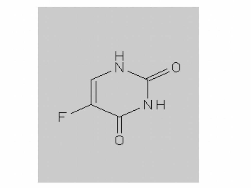
Interference with glycosyltransferases altering cell membranes (Fig. 2).
It has been proved in laboratory tests that 5 FU produces a slight reduction of collagensynthesis in normal fibroblasts but a drastic reduction in altered ones, as in Dupuytrenillness[3]. Apparently it also inhibits the collagen synthesis stimulation effect of TGF1(transforming growth factor).
5 FU has been used for years in treatment of premalignant and malignant lesions ofskin and mucosa due to its selective toxicity for dysplastic epithelium and fibroblasts.
The first application of 5 FU was as an antifibrotic to prevent fibrous scarring afterglaucoma surgery and to avoid relapse in pterigion surgery in the sixties[4, 5, 6, 7, 8,9]. In February of 1999 its use was reported to prevent fibrous adherence after tendonreconstructive surgery. One month after Fitzpatrick published his seven yearexperience of over a thousand patients with 5 FU in hypertrophic scars and keloids.
This magnificent work encourage our team to include it in our office with positiveresults. Since Lambros published his work in 2004 [10] our team also addedhyaluronidase. Fig.2- Molecular structure of 5 FU
This last is a enzyme that increases connective tissue permeability by hyaluronic acidhydrolysis. Hyaluronic acid is a polysaccharide of connective tissue and other

specialized tissues as the umbilical cord and the vitreous humor. Hydrolysis is donebetween the C1of glucosamine and C4 of glucuronic acid. This reduces temporarily theintercelular cement viscosity, promoting diffusion of injected solution, exudates andtransudates facilitating absorption.
Triamcinolone is a steroid, it diffuses through cell membranes binding withcytoplasmic receptors that are translocated to the nucleus generating the transcriptionof proteins that are responsible of their effects. It reduces tissue response toinflammation, reducing it symptoms without treating the specific cause. To do this itreduces the white blood cells (WBC) migration to the affected tissue. The mostimportant effects are:
Inhibition of phagocytosis.
Inhibition of liberation of lysosomal enzymes and inflammatory mediators.
Reduction of capillary permeability and WBC adhesion to capillary endothelium.
Reduction of blood concentration of T-cells, eosinophils and monocytes.
Reduction of immunoglobulin binding with cellular receptors.
In the past 14 years we have been using a preparation of 5FU, hyaluronidase andtriamcinolone for the nasal area and we have not had the complications and side effectsobserved with steroids monotreatment.
5 FU (Fluorouracil- Filaxis 500mg) is presented commercially as ampoules of 10
ml containing 50 mg per ml. Triamcinolone (Kenacort-A-BSM) is used in its acetonideform of 40mg/ml, and is commercialized in ampoules of 1ml (Fig. 3). Hyluronidase(Unidasa Roux Ocefa) is commercialized in ampoules that
Fig.3- Commercial presentations of 5 FU and triamcinolone
contains 500 UI of testicular ovine freeze dried powder hyaluronidase. We use2.7ml of 5 FU and 0.3ml of triamcinolone to reconstitute hyaluronidase (5 FU
solution). For the application a 0.3 or 0.5 ml Luer lock syringe and 30G needleare preferred. This allows a better dosage and correct plane of infiltration. Wehad never used more that 0.5 ml of the solution in the nasal area per session.
The infiltration is done with multiple punctures in the "heart" of the fibroticlesion. The first month one session is done per week, then is spaced to everyfifteen days. The improvement is evaluated with 3 parameters: hardeningreduction, loss of volume and reduction of cutaneous retraction. Positivechanges are observed since the first session, not only in the above parametersbut also in aesthetics and pain- itching symptomatology. Some cases arecomplemented with kinesiology treatment in order to obtain a better functionaland cosmetic result.
If after finishing the treatment a surgical procedure was needed to improve theresult, the surgical piece was send to the pathologist and compared to nontreated scarring tissue resections. Results informed reduction of fibrosis andrearrangement of collagen tissue in the treated pieces. These results areillustrated in the images. In the tissue of the right side of the scar proliferationof fibrous tissue, great amount of fibroblasts and collagen fibers formingtangles is seen (Fig. 4). In the tissue of the left, treated with 5 FU solution, thereare less fibroblasts, and the collagen fibers adopt a parallel disposition (Fig. 5).
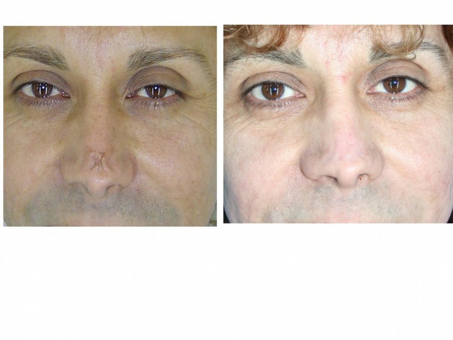
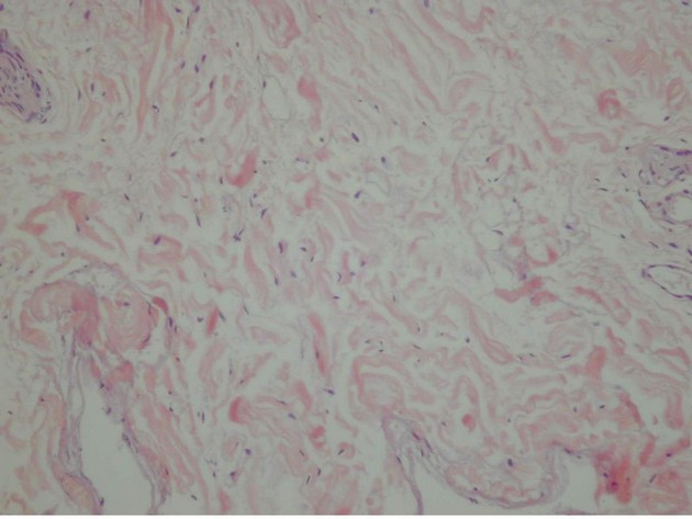
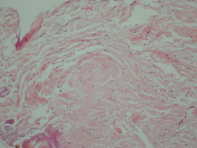
Fig.4- Microscopic image of fibrosis without any infiltration treatment(upper)
Fig.5- Microscopic image of fibrosis after infiltration with 5 FU(lower)
CLINICAL APPLICATIONS
Hypertrophic scars or keloids fixed to deep planes
"Supratip" post rhinoplasty fibrosis
Post rhinoplasty fibrosis (as preparation for other treatments)
Foreign body granulomas
Post burn retractions
Hypertrophic scars or keloids fixed to deep planes
The nose, being a mid facial and projected structure, is exposed to trauma
that leave scarring sequels in the skin . Traumatic scarring if healed by
second intention generates unaesthetic scars. The use of 5 FU,
triamcinolone and hyaluronidase for hypertrophic scars and keloids in the
nasal area is in our hands more effective and safe that the use of steroids
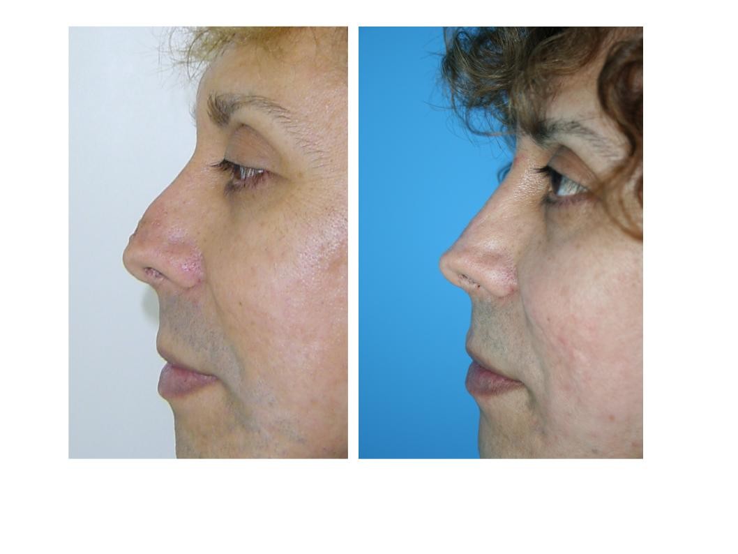
Fig.6- Nasal tip scar after cartillage graft infection and extrusion. Frontal
view before and after 5FU injection and dermabration.
alone. When this scars are fixed to deep planes, the infiltration is done as
preparation to subcision and then a filler as Polymethylmetacrilate
(PMMA) is used to prevent the relapse of the adherence.
"Supratip" post rhinoplasty fibrosis
Fig.7- Nasal tip scar after cartillage graft infection and extrusion. Lateral view before and after 5FU
injection and dermabration.
One of the most frequent complications of rhinoplasty is the healing fibrosisthat is formed over the time known as "fibrous supratip". It can be the result ofa bad executed rhinoplasty or defective healing process. Is very frequent amongbeginners and even experts still have this complication. There are many causesof supratip deformity and each demand a specific treatment. Bahman Guyuron[11] conducted a clinical and histological study to unmask the surgical causes ofthis deformity. This study shows that clinical supratip is observed in 9% ofprimary and 36% of secondary consults of rhinoplasty. In primary cases thedeformity is the result of: tip inadequate projection, caudal dorsum overprojection, lateral inferior cartilages cephalic orientation, or a combination ofthese. In secondary cases the deformity is the result of: sub correction orovercorrection of caudal dorsum, over resection of medial valve, sub projected
tip, or a combination of these. The histophatological study of the supratip softtissue demonstrated significant fibrosis in 14 from 16 secondary patients and 13of 23 primary patients. The supratip deformity can be avoided throughappropriate resection of caudal dorsum (in order not to leave dead space), nasaltip projection and joining with stitches the subcutaneous tissue over thecartilages in the supratip area. When early diagnosed, if the tip has adequateprojection and the supratip tissue can be collapsed by pressure, the electivetreatment is compressive tape. If after 6 weeks the response is not positive 0.2to 0.4 ml of 5 FU solution is injected in the deep (Fig 8 and 9)
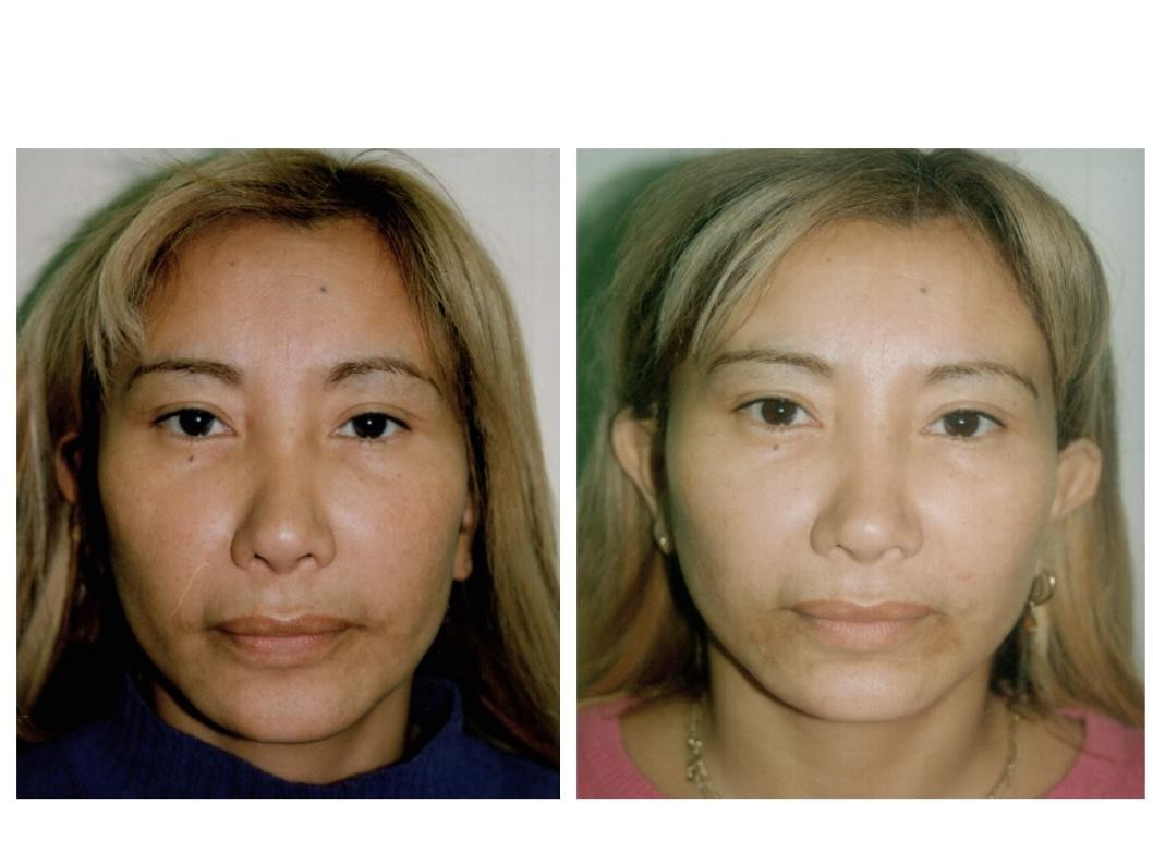
Fig.8-Fibrose supratip after previous procedure solve after 5 year with 0.3 ml of 5FU single injection.
Frontal view.
subcutaneous tissue. This infiltration can be repeated monthly until reaching thedesired result (The judicious use 5 FU solution can help in most supratip Fig.9-Fibrose supratip after previous procedure solve after 5 year with 0.3 ml of 5FUsingle injection. Frontal view. Lateral view.
deformities caused by fibrosis when the caudal septum and the tip cartilages arestrong. If the cartilaginous frame is weak, infiltration will not solve theproblem.Infiltration can reduce a big supratip to a small one, but with animportant risk of skin atrophy.
Other authors as Gruber [12] prefer to start with infiltration after 4 to 6 weeksof surgery, when most edema has disappeared. He uses 1 to 2 mg oftriamcinolone (0.1 to 0.2cc of triamcinolone 10mg /cc) in the supratip or otherfibrous area. Pastorek [13]uses small steroids dose during and immediately aftersurgery having excellent results in his hands. If a severe supratip deformity isthe consequence of inadequate cartilage resection or sub projection of the tip, asurgical correction or bioplasty are needed. Sheen[14] suggested in 1979 thatmost supratip deformities were caused by caudal dorsum over resection. Heproposed then the use of cartilage graft to correct this deformity.
Nowadays it is widely accepted that healing fibrotic tissue formed to fill thedead space is the most frequent cause of supratip deformity post rhinoplasty.
But it was not until Guyuron's work that this was scientifically confirmed.
Fig. 10- Patient with 3 previous rhinoplasties. Three aplications of 5 FU solution were done.
Post rhinoplasty fibrosis, as preparation for other treatments
After several surgical procedures, the nose can be involved in different grades offibrosis turning the skin hard and inelastic. Before performing a secondary rhinoplastyor a bioplasty we prefer preparing the area with some applications of the 5 FU solutionin order to soften the tissue (Fig. 10).
Foreign body granulomas
The use of fillers in the nasal area is a very popular method to correct slightdeformities. There are many different materials and each of them produce a differentdegree of fibrotic reaction in the tissue. In most cases, this is a controlled reaction andlead to the expected result. But in some cases, an over reaction of patient immunesystem, bad application technique or the chemistry of the material used, provoke theformation of foreign body granulomas, resulting in unaesthetic deformities.
The use of triamcinolone in this cases can generate skin atrophy, thus turning thedeformity more visible. When the filler used is hyaluronic acid, pure hyaluronidaseshould be infiltrated as stated by Brody [15]and Hirsch [16]. Infiltration is done with100 UI in the area of hyaluronic acid excess (Fig. 11). This dissolves the acid, solvingthe fibrosis. When alloplastic materials were used, we preferred the 5 FU solution forinfiltration because it does not have the side effects of steroids monotreatment. Theapplication has a double effect, the pharmacological effect of the drugs, and themechanical effect of disruption by the fluid pressure
Post burn retractions
The nasal skin usually gets affected in facial burns. The scarring retraction can deformthe tip, ala or both, stretching the nostrils. We use the 5 FU solution infiltration in thesefibrous areas in order to increase the tissue elasticity and prepare them for secondaryprocedures as PMMA bioplasty or compound auricular graft.
The use of 5 FU solution, in young patients mostly, can cause tissue necrosis. This isrelatively frequent in keloid treatment, resulting in ulcers or brown stains in theapplication spots. However in the nasal region we have not observed this complication,mainly because of the low dosage. In this way, by using progressive treatment insteadof aggressive one, side effects are kept to minimum.
Through this chapter we presented our experience in the treatment of scars, fibrosis,retractions and deformities over the nasal skin. The results of our 14 year experience
with 5 FU solution ( 5 FU, triamcinolone and hyaluronidase) has the histopathologicaldemonstration of effectiveness in collagen fibers synthesis reduction and fiberrearrangement, thus explaining the positive clinical effect
CONFLICT OF INTEREST
Fitzpatrick R. Treatment of inflamed hypertrophic scars using intralesional 5-FU.
Dermatol. Surg.1999 ;25(3):224-232.
Blugerman G. Keloids and Hipertrofic scars. Disertation. Congreso de Cirugía Plástica
del Cono Sur, Porto Alegre, Brasil, Mayo 2001
Jemec, B., C. Linge, et al. The effect of 5-fluorouracil on Dupuytren fibroblast
proliferation and differentiation. Chir Main 2000;19(1):15-22
Blugerman G, Schavelzon D. Intralesional use of 5-FU in subcutaneous fibrosis. J
Drugs Dermatol 2003; 2: 169-171
Yamamoto T, Varani J, Soong HK, Lichter PR.: Effect of 5-fluorouracil and mitomycin
on cultured rabbit subconjunctival fibroblast. Ophthalmology 1990;97:1202-1210.
Yeatts RP, Ford JG, Stanton CA, Reed JW. Topical 5-fluorouracil in the intra epithelial
neoplasia of the conjuntiva and cornea. Ophthalmology 1995; 102:1388-44.
Lee DA, Shapourifar-Tehrani S, Kitada S. The effect of 5-fluorouracil and cytarabine
on human fibroblasts from Tenon's capsule. Invest Ophthalmol Vis Sci 1990;31:1848-55.
Schellini Silvana A. Uso do 5-fluorouracil no intra-operatório da cirurgia do pterígio.
Arq. Bras. Oftalmol. 2000 63(2) 111-114
Acali Augustine et all. Decrease in adhesión formation by a single application of
5Fluorouracil after flexor tendon injury. Plast. Reconstr Surg.1999;103(1):151-158
Lambros V. The use of hyaluronidase to reverse the effects of hyaluronic acid filler.
Plast. Reconstr. Surg. 2004 ; 114(1):277
Guyuron, Bahman, Supratip Deformity: A Closer Look. Plast. Reconstr. Surg.
2000.105(3):1140-1151,
Gruber, Ronald P. M.D. Supratip Deformity: A Closer Look by Bahman Guyuron,
M.D., Louis DeLuca, M.D., and Richard Lash, M.D. Plast. Reconstr. Surg. 2000 105(3):1152-1153,
Pastorek, N.Surgery of the Nasal Tip. Disertation. Dallas Rhinoplasty Symposium,
Dallas, Texas, March, 1999.
Sheen, J. H. A new look at supratip deformity. Ann. Plast. Surg. 1979 ;3(6):498-504.
Brody, H.J. The use of hyaluronidase in the treatment of granulomatous hyaluronic
acid reactions or unwanted hyalronic acid misplacement. Dermatol. Surg. 2005 ;31(8 Pt 1):893-7.
Hirsch R.J. Correcting superficially placed of Hyaluronic Acid. Skin and Aging. 2007;
15 (1) 36-38.
Source: http://ijcs.org/49/104/1089/5%20Fluorouracil%20hialuronidase%20and%20triamcinolon%20in%20the%20nasal%20area%20blugerman%20at%20al.pdf
Our mission is to help the people of Canada maintain and improve their health. Health Canada Additional copies are available from: Telephone: (613) 954-5995 Fax: (613) 941-5366 This publication is also available on the Canada's Drug Strategy Division web site at the following address: It can be made available in/on computer diskette/large print/ audio cassette/braille, upon request.
a-se- feks DESCRIPTIONThe active ingredient in ACIPHEX® Delayed-Release Tablets is rabeprazole sodium, a substituted benzimidazole thatinhibits gastric acid secretion. Rabeprazole sodium is known chemically as 2-[[[4-(3-methoxypropoxy)-3-methyl-2-pyridinyl]-methyl]sulfinyl]-1H–benzimidazole sodium salt. It has an empirical formula of C18H20N3NaO3S and a molecularweight of 381.43. Rabeprazole sodium is a white to slightly yellowish-white solid. It is very soluble in water andmethanol, freely soluble in ethanol, chloroform and ethyl acetate and insoluble in ether and n-hexane. The stability ofrabeprazole sodium is a function of pH; it is rapidly degraded in acid media, and is more stable under alkalineconditions. The structural formula is:


