Rosiglitazone prevents nutritional fibrosis and steatohepatitis in mice
Scandinavian Journal of Gastroenterology, 2009; 44: 358365
Rosiglitazone prevents nutritional fibrosis and steatohepatitis in mice
YUE-MIN NAN1, NA FU1, WEN-JUAN WU1, BAO-LI LIANG1, RONG-QI WANG1,SU-XIAN ZHAO1, JING-MIN ZHAO2 & JUN YU3,4
1Department of Traditional and Western Medical Hepatology, the Third Hospital of Hebei Medical University, Shijiazhuang,China, 2Department of Pathology, Beijing 302 Hospital, Beijing, China, 3Institute of Digestive Disease and Li Ka ShingInstitute of Health Sciences, Department of Medicine and Therapeutics, The Chinese University of Hong Kong, Hong Kong,and 4Department of Gastroenterology, the First Hospital of Hebei Medical University, Shijiazhuang, China
AbstractObjective. Currently, no agent has been confirmed as preventing the fibrosing progression of non-alcoholic steatohepatitis(NASH). In this study, rosiglitazone was used in the clinical treatment of insulin resistance in patients with type 2 diabetesmellitus. However, its protective effect on non-alchoholic fibrosing steatohepatitis is not clear. The study aimed to elucidatethe effect and the mechanism of rosiglitazone in inhibiting nutrition-related fibrosis in mice. Methods. C57BL6/J mice werefed a high fat, methionine-choline deficient (MCD) diet for 8 weeks to induce hepatic fibrosis, and rosiglitazone was given inthe treated group. The effect of rosiglitazone was assessed by comparing the severity of hepatic fibrosis in liver sections, theactivation of hepatic stellate cells (HSCs) and the expression of TGF-b1 and connective tissue growth factor (CTGF).
Results. At week 8, MCD-diet-induced fibrosing NASH models showed increased serum ALT and AST levels, severehepatic steatosis, and infiltration of inflammation and fibrosis which, associated with down-regulated PPARg mRNA andprotein expression, up-regulated a-SMA protein expression and enhanced TGF-b1, CTGF mRNA and protein expression.
Rosiglitazone significantly lowered serum ALT and AST and it reduced MCD-induced fibrosis by repressing levels of
For personal use only.
a-SMA protein expression and pro-fibrosis factors TGF-b1 and CTGF. It also restored expression of PPARg.
Conclusions. The present study provides clear morphological and molecular biological evidence of the protective roleof rosiglitazone in ameliorating nutritional fibrosing steatohepatitis. Rosiglitazone may ameliorate hepatic fibrosis byactivating PPARg, which can inhibit HSC activation and suppress TGF-b1 and CTGF expression.
Key Words: Fibrosing steatohepatitis, hepatic stellate cells, non-alcoholic steatohepatitis, peroxisome proliferator activatedreceptor gamma, rosiglitazone
liferation, loss of vitamin A storing capability,expression of a-smooth muscle actin (a-SMA) and
The pathological progression of non-alcoholic fatty
Scand J Gastroenterol Downloaded from informahealthcare.com by Nyu Medical Center on 06/25/10
overproduction of ECM components. Among profi-
liver disease (NAFLD) includes simple hepatic
brogenic cytokines, transforming growth factor beta
steatosis, steatohepatitis, hepatic fibrosis and cirrho-sis. Up to now, the pathogenesis of non-alcoholic
1 (TGF-b1) and its downstream effective factor,
steatohepatitis with hepatic fibrosis remains unclear.
connective tissue growth factor (CTGF), are the
Hepatic stellate cells (HSCs) are the primary source
master factors for promoting HSC activation, ECM
of excessive production of extracellular matrix
synthesis and secretion of other profibrogenic factors
(ECM), and activation of HSCs is the critical event
in hepatic fibrosis [1]. HSCs belong to the non-
Peroxisome-proliferator-activated receptor gamma
parenchymal cells in Disse space. Many profibro-
(PPARg) is a soluble transcription factor. Recent
studies have demonstrated that PPARg expres-
into activated HSCs which transdifferentiate to a
sion decreases markedly with the activation of
myofibroblast phenotype characterized by cell pro-
HSCs, which indicates that PPARg is prominent in
Correspondence: Yue-min Nan, Traditional and Western Medical, Department of Hepatology, Third Hospital of Hebei Medical University, Shijiazhuang,China. Tel: 86 311 88602151. Fax: 86 311 87023626. E-mail:
[email protected]
(Received 9 August 2008; accepted 1 October 2008)
ISSN 0036-5521 print/ISSN 1502-7708 online # 2009 Informa UK Ltd.
DOI: 10.1080/00365520802530861
Rosiglitazone and fibrosing steatohepatitis
maintaining the quiescent phenotype of HSCs [5].
hepatic steatosis, necroinflammation and fibrosis in
This implies that PPARg may be a potential target in
accordance with the 2006 Guidelines for Diagnosis
preventing hepatic fibrogenesis and the selective
and Treatment of Non-Alcoholic Fatty Liver Dis-
PPARg agonist may be used as a therapeutic agent
eases [7] and the 2001 programme of prevention and
for liver fibrosis. Rosiglitazone, an agonist of PPARg,
cure for viral hepatitis [8] issued by the Chinese
may protect the pathogenesis and progression of
Society of Hepatology of the Chinese Medical
fibrotic disease by activating PPARg. The aim of this
study was to investigate the effect of rosiglitazoneon activation of HSCs and expression of PPARg,TGF-b1 and CTGF in mice with hepatic fibrosis
induced by the MCD diet [6].
Immunostaining for CTGF and a-SMA was per-formed in paraffin-embedded liver sections using thespecific antibodies (Santa Cruz, Calif., USA) and an
Material and methods
avidin-biotin complex (ABC) immunoperoxidase
Animals and treatments
method. Briefly, endogenous peroxidase activitywas blocked by treating sections with 3% hydrogen
Eight-week-old male C57BL6/J mice with body
peroxide. After blocking with 10% non-immunized
weights between 20 and 25 g were obtained from
goat serum, the primary specific antibodies for
the Experimental Animal Center of the Chinese
CTGF (dilution 1:200) and for a-SMA (dilution
Academy of Medical Sciences and were housed in a
1:200) were applied. Primary antibodies were
228C-controlled room under a 12-h lightdark
omitted and non-immunized goat serum was used
cycle. They had free access to water and were
for negative controls. After extensive rinsing, the
allowed to adapt to their food and environment for
biotinylated secondary antibody and ABC complex
1 week before the start of the experiment. The
/HRP were applied. Peroxidase activity was visua-
C57BL6/J mice were divided into 3 groups (10
lized by applying diaminobenzidine to the sections,
/group) and fed a control diet (ICN, Aurora, Oh.,
which were then counter-stained with haematoxylin.
USA), a methionine and choline deficient (MCD)
Quantitative analysis of CTGF and a-SMA-stained
diet (ICN) or an MCD diet supplemented with
liver sections was performed by morphometric
rosiglitazone (50 mg kg1 d1). During the experi-
ments, their body weights and rate of diet con-sumption were recorded. After 8 weeks, all of the
For personal use only.
animals were killed and blood collected from the
Determination of hepatic PPARg, TGF-b1 and CTGF
femoral artery. Livers were weighed and fixed in
10% formalin for histological analysis or snap-frozen in lipid nitrogen followed by storage at
Total RNA was isolated with TRIzol reagent
808C in a freezer until required. All the protocols
(Saibaisheng Biological Technique Institute, Beijing,
and procedures were carried out following the
China). For RT-PCR, 5 mg total RNA was reverse-
guidelines of the Hebei committee for care and
transcribed with M-MLV in accordance with the
use of laboratory animals, and were approved by
manufacturer's instructions. cDNAs were amplified
the Animal Experimentation Ethics Committee of
using specific sets of primers for PPARg (sense,
the Hebei Medical University.
Scand J Gastroenterol Downloaded from informahealthcare.com by Nyu Medical Center on 06/25/10
gaggcctg-3?), TGF-b1 (sense, 5?-caacgccatctatga-gaaaacc-3?;
Measurement of serum ALT and AST
c-3?) and CTGF (sense, 5?-caaagcagctgcaaatacca;
Serum alanine aminotransferase (ALT) and aspar-
antisense, 5?-ggccaaatgtgtcttccagt). The PCR proce-
tate aminotransferase (AST) levels were determined
dure for PPARg consisted of 30 cycles of denatura-
using spectrophotometric assay kits (Sigma, St.
tion at 948C for 45 s, annealing at 578C for 45 s,
Louis, Mo., USA) with an automatic biochemical
extension at 728C for 5 min, with initial denatura-
analyser (Olympus UA2700, Japan).
tion of sample cDNAs at 948C for 5 min before PCRand an additional extension period of 10 minafter the last cycle. The different procedure for
Histological examination
TGF-b1 and CTGF is annealing at 608C for 45 s.
The fat on the frozen liver sections (5 mm thick) was
In parallel, PCR reactions were performed with
detected with Sudan IV stain, a conventional
primers coding for the housekeeping gene b-actin
method for fatty tissue in histology. Haematoxylin
and eosin-stained paraffin-embedded liver tissues
5?-gctgatccacatctgctggaa-3?) to control for equal
isolated from mice (5 mm thick) were graded for
amounts of template cDNAs. A quantity of 6 ml of
Y.-M. Nan et al.
PCR products was analysed in an 8% sodium
Effect of rosiglitazone on liver inflammatory injury
phoresis (SDS-PAGE) with a 100-bp DNA marker.
Densitometric analysis of PCR products was per-
As indicated in Table 1, mice fed the MCD diet
formed by computer software and standardized by
showed significantly higher serum ALT and AST
levels compared with control mice, indicating hepa-tic injury ( pB0.05), and a significant reduction wasnoted after rosiglitazone treatment ( pB0.05). How-
Western blot analysis of hepatic proteins of PPARg and
ever, serum triglyceride and cholesterol concentra-
tions were not significantly altered.
Hepatic tissues (10% w/vol) were homogenized inlysis buffer containing 20 mM Tris-HCl (pH 7.4),
Effect of rosiglitazone on liver histology
1% Triton X-100, 140 mM NaCl, 1 mM phenyl-methylsulfonyl fluoride (PMSF) and aprotinin, leu-
Compared to the controls (Figure 1A and 2A), the
peptin, soybean trypsin inhibitors (1 mg/ml each).
liver section from mice fed the MCD diet exhibited
Total protein was extracted and concentration was
disordered lobule structure, macrosteatosis in Zone
measured by the Bradford method (DC protein
3, spot or focal hepatocyte necrosis and inflamma-
assay; Bio-Rad, Hercules, Calif., USA) as previously
tory infiltration (Figure 1B) and portal and perisi-
described [9]. A quantity of 100 mg protein was
nusoidal fibrosis (Figure 2B). Treatment with
separated by 10% sodium dodecyl sulphate-poly-
rosiglitazone markedly improved hepatic steatosis,
acrylamide gel electrophoresis and then transferred
inflammatory infiltration (Figure 1C) and fibrosis
onto equilibrated polyvinylidene difluoride mem-
(Figure 2C).
brane (Amersham Biosciences, Buckinghamshire,UK) by electroblotting. Membranes were blocked
Effect of rosiglitazone on body weight and liver weight
with 5% skim milk in 1 TBST for 1 h, then
incubated with specific antibodies against PPARg,TGF-b1 or b-actin (Santa Cruz Biotechnology,
Similar to what has been reported previously [10],
Santa Cruz, Calif., USA) overnight at 48C. After
administration of the MCD diet caused body weight
incubation with secondary antibody, proteins were
loss and liver weight gain (Table 2). Administering
For personal use only.
detected by enhanced chemiluminescence (ECL;
rosiglitazone significantly increased body weight by
up to 7% ( pB0.05) under the MCD diet andreduced liver weight ( pB0.05).
Statistical analysis
Effect of rosiglitazone treatment on CTGF expression
The Statistical Package for Social Science software(v. 13.0; SPSS Inc., Chicago, Ill., USA) was used for
As shown in Figure 3A, CTGF, a cysteine-rich,
statistical analysis. The data are presented as
means9SD. Statistical analysis was carried out by
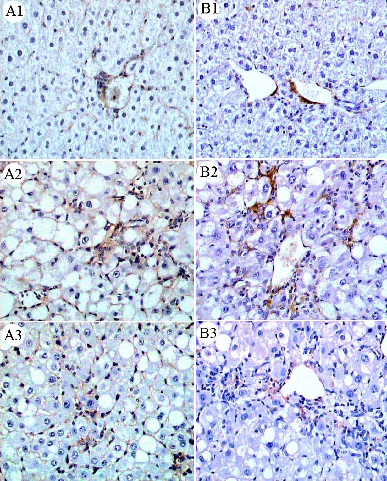
dramatically increased mostly in fibrous tissue (Fig-
one-way analysis of variance (ANOVA) and the
ure 3A2) compared to CTGF in the control mice
Scand J Gastroenterol Downloaded from informahealthcare.com by Nyu Medical Center on 06/25/10
StudentNewmanKeuls test (for evaluating differ-
(Figure 3A1), but was significantly reduced by
ences between groups). A p-value of B0.05 was
rosiglitazone treatment (Figure 3A3) as determined
considered statistically significant.
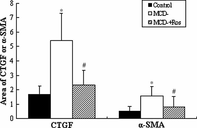
by a computerized morphometric analysis (Figure 4).
Table 1. The effect of MCD diet with or without rosiglitazone on serum characteristics
Triglyceride (mmol/L)
Cholesterol (mmol/L)
NOTE. Data are mean9SD ( n10/group).
*PB0.05 relative to mice fed the control diet.
#PB0.05 relative to mice fed the MCD diet.
Rosiglitazone and fibrosing steatohepatitis
Figure 1. The effect of rosiglitazone on the liver histology in MCD-diet-induced fibrosing steatohepatitis. Haematoxylin and eosin-stainedliver sections from mice fed: (A) the control diet, (B) the MCD diet, (C) the MCD diet supplemented with rosiglitazone (50 mg kg1 d1)(black arrows indicate hepatic steatosis). Experimental duration is 8 weeks. Slides are representative of 10 animals per group.
Figure 2. The effect of rosiglitazone on hepatic fibrosis in mice fed the MCD diet. Masson-trichrome staining for collagen fibres in liversections from mice fed: (A) the control diet, (B) the MCD diet, (C) the MCD diet supplemented with rosiglitazone (50 mg kg1 d1)(black arrows indicate hepatic fibrosis). Experimental duration is 8 weeks. Slides are representative of 10 animals per group.
For personal use only.
Effect of rosiglitazone treatment on HSC activation
with that in mice fed the control diet. Rosiglitazonerestored the expression of PPARg (Figure 5). The
To provide further evidence of the anti-fibrotic
mRNA and protein expression of TGFb1 was
property of rosiglitazone in MCD feeding mice, we
increased in MCD-feeding mice and was signifi-
determine the effects of rosiglitazone on hepatic
cantly blunted by rosiglitazone treatment (Figure 5).
a-SMA protein expression, a well-established marker
Consistent with the protein expression determined
of HSC activation during liver fibrogenesis. As
by immunohistochemistry (Figure 3A), treatment
shown in Figure 3B and 4, and as compared to
with rosiglitazone significantly down-regulated the
MCD mice, rosiglitazone significantly suppressed
mRNA expression of CTGF induced by MCD
a-SMA expression.
feeding (Figure 5).
Scand J Gastroenterol Downloaded from informahealthcare.com by Nyu Medical Center on 06/25/10
Analysis of transcripts and proteins of genes associated
with HSC activation and liver fibrosis
A high fat, methionine-choline-deficient diet in
Both mRNA and protein expression of PPARg were
mice results in hepatic steatosis, inflammation
dramatically decreased in mice fed MCD compared
and fibrosis. This is distinct from the high fat
Table 2. Weight changes in mice fed the control or MCD diet with or without Rosiglitazone
Number of animals
Body weight (% change)
Relative liver weight (% of body weight)
NOTE. Data are mean9SD.
*PB0.05 relative to mice fed the control diet.
#PB0.05 relative to mice fed the MCD diet.
Y.-M. Nan et al.
For personal use only.
Scand J Gastroenterol Downloaded from informahealthcare.com by Nyu Medical Center on 06/25/10
Figure 3. Effect of rosiglitazone on protein expression of CTGF and a-SMA. (A) Immunostaining for CTGF protein in (A1) mice fed thecontrol diet, (A2) mice fed the MCD diet and (A3) mice fed the MCD diet supplemented with rosiglitazone 50 mg kg1 d1. (B)Immunostaining for a-SMA protein in (B1) mice fed the control diet, (B2) mice fed the MCD diet and (B3) mice fed the MCD dietsupplemented with rosiglitazone 50 mg kg1 d1. There were inductions in CTGF and a-SMA staining in mice fed MCD for 8 weeks, andadministering rosiglitazone blunted the induction of both CTGF and a-SMA.
diet-induced liver injury, but resembles the hepatic
steatohepatitis with liver fibrosis, resulting in char-
pathohistological alteration in human non-alcoholic
acteristic pathology of steatosis, mixed inflammatory
steatohepatitis. We used this representative experi-
cell infiltration hepatocyte necrosis, and fibrosis
mental model of fibrosing steatohepatitis to investi-
in the pericellular, perisinusoidal and portal area.
gate the mechanism behind rosiglitazone preventing
Concomitant with increased levels of serum ALT and
the progress of hepatic fibrosis.
AST, administering rosiglitazone in MCD-fed mice
Following the MCD diet for 8 weeks, mice rapidly
resulted in attenuation of the fibrosing steatohepati-
and consistently developed a severe pattern of
tis, as evidenced by decreased ALT and AST levels
Rosiglitazone and fibrosing steatohepatitis
CCl4 and bile duct ligation (BDL) in murine. Ourresults were also supported by the evidence thatrosiglitazone protected
induced steatohepatitis [12] and patients withNASH with reduced inflammation and liver injury[13,14]. Restoration of the body weight of mice fedthe MCD diet treated with rosiglitazone, with areduction of liver weight and hepatic fat contentand serum ALT level, suggests a shipment of fat fromthe liver to adipose tissue. The roles of adipose tissueand its PPARg on hepatic fat accumulation have beendemonstrated previously in loss-of-function studies,where it has been shown that genetically engineered
Figure 4. Effect of rosiglitazone on quantitative protein expres-sion of CTGF and a-SMA. The expressions of CTGF and
mice with no adipose tissue or adipose-tissue-specific
a-SMA were estimated by average area density (areas of positive
PPARg knockout automatically acquired fatty liver
cells/total areas 100%) in each scope (200-fold) to observe
[1517]. On the other hand, activation of PPARg by
10 scopes at every section (2G). Data are shown as mean9SD
rosiglitazone promotes lipogenesis in adipose tissue.
( n10/group), *PB0.05 compared with control mice, #PB0.05
Besides enhancing fat redistribution, the ameliorat-
rosiglitazone-treated MCD-fed compared with the mice fed theMCD diet.
ing effect of rosiglitazone was also mediated throughmodulating adipokines. Other than inflammatory
and diminished histologic evidence of fibrosis.
cytokines in liver induced by lipotoxicity of the
In keeping with our finding, Galli et al. [11]
accumulated fat, adiponectin derived from adipose
tissue certainly plays an anti-inflammatory role in the
liver fibrosis induced by dimethyl nitroxide (DMN),
progression of steatohepatitis [18].
For personal use only.
Scand J Gastroenterol Downloaded from informahealthcare.com by Nyu Medical Center on 06/25/10
Figure 5. Effects of rosiglitazone on hepatic PPARg, TGF-b1 and CTGF expression in MCD feeding mice. (A) PPARg, TGF-b1 andCTGF mRNA expression was analysed by RT-PCR. The level of PPARg was low and TGF-b1 and CTGF were high in MCD mice.
However, rosiglitazone could restore PPARg and suppress TGF-b1 and CTGF expression. The expression of mRNA was estimated by ratioagainst b-actin. (B) Protein expression of PPARg and TGF-b1 was determined by Western blot. The changes in protein were consistent withthose of the mRNA. Western blots were scanned by densitometry and the data presented as relative intensity units against b-actin. Each barrepresents the mean9SD of four to seven mice. *PB0.05 compared with control mice, #PB0.05, ##PB0.01, ###PB0.001 rosiglitazone-treated MCD-fed compared with the mice fed the MCD diet
Y.-M. Nan et al.
HSC activation is key in the early phase of liver
mechanisms of its action could be involved in
fibrosis and activated HSC is accompanied by a high
activating PPARg to reverse activated HSCs into a
expression of a-SMA protein. Many kinds of fibro-
quiescent phenotype and reduce the expression of
genic cytokines are involved in HSC activation in
TGF-b1 and CTGF.
keeping HSCs quiescent [5]. We found that reducedexpression of PPARg and enhanced expression of
a-SMA in fibrosing steatohepatitis in mice fed MCDand rosiglitazone could dramatically up-regulate
This work was supported financially by the Wang
PPARg and down-regulate a-SMA. Thus, rosiglita-
zone might delay or inhibit the progression of liver
20070021 and by a Research Grants Council
fibrosis through agitating PPARg, which might
Competitive Earmarked Research Grant (CUHK
be the major mechanism of anti-fibrogenesis in
non-alcoholic fatty liver disease.
The important role of TGF-b1 in liver fibrosis has
Declaration of interest: The authors report no
been well documented [1922]. It has been pro-
conflicts of interest. The authors alone are respon-
posed that TGF-b1 secreted by fibroblasts, Kupf-
sible for the content and writing of the paper.
fer cells and T-lymphocytes [23] is a key mediatorin the pathogenesis of liver fibrosis, and has a varietyof biological effects that include activation of HSCs
[24,25]. It could inhibit secretion of matrix metallo-
[1] Uemura M, Swenson ES, Gac¸a MD, Giordano FJ, Reiss M,
proteinases and promote tissue inhibitor of metallo-
Wells RG. Smad2 and Smad3 play different roles in rat
proteinases, and it could up-regulate platelet-derived
hepatic stellate cell function and alpha-smooth muscle actin
growth factor receptor, all of which are involved in
organization. Mol Biol Cell 2005;16:421424.
hepatic fibrogenesis. Our study reveals that the
[2] Tomita K, Tamiya G, Ando S, Ohsumi K, Chiyo T, Mizutani
A, et al. Tumor necrosis factor a signaling through activation
expression of TGF-b1 is significantly increased in
of Kupffer cells plays an essential role in liver fibrosis of non-
fibrosing steatohepatitis in mice fed MCD, and is
alcoholic steatohepatitis in mice. Gut 2006;55:41524.
suppressed by treatment with rosiglitazone. This
[3] Friedman SL. Molecular regulation of hepatic fibrosis: an
effect might take place at the early stage of
integrated cellular response to tissue injury. J Biol Chem
HSCs activation [3,26], as activated HSCs secrete
[4] Gressner OA, Lahme B, Demirci I, Gressner AM, Weis-
TGF-b1, which, combined with its receptor through
kirchen R. Differential effects of TGF-beta on connective
For personal use only.
autocrine or paracrine in turn, activates HSCs and
tissue growth factor (CTGF/CCN2) expression in hepatic
stellate cells and hepatocytes. J Hepatol 2007;47:699710.
PPARg to reverse activate HSCs into a quiescent
[5] Hazra S, Miyahara T, Rippe RA, Tsukamoto H. PPAR
type that reduces the expression of TGF-b1. As
gamma and hepatic stellate cells. Comp Hepatol 2004;3
another fibrogenesis factor, CTGF is the down-
[6] Nan Y, Wu W, Yao X, Wang L. The role of apoptosis and the
stream effective factor of TGF-b1 [27,28]. We found
related genes in non-alcoholic steatohepatitis. Zhonghua
that CTGF expression was consistent with TGF-b1
Gan Zang Bing Za Zhi 2007;15:416.
expression. Rosiglitazone could also inhibit CTGF
[7] Fatty liver and Alcoholic Liver Disease Study Group of the
expression in fibrosing steatohepatitis in mice fed
Chinese Liver Disease Association. Guidelines for diagnosisand treatment of nonalcoholic fatty liver diseases. Zhanghua
MCD. Several studies have shown that leptin exerts
Ganzangbing Zazhi 2006;14:1613.
a pro-fibrogenic activity that acts directly on hepatic
Scand J Gastroenterol Downloaded from informahealthcare.com by Nyu Medical Center on 06/25/10
[8] Chinese Society of Infectious Diseases Study and Parasitol-
stellate cells [29,30] to increase expression of
ogy and Chinese Society of Hepatology of Chinese Medical
TGF-b1 and a-SMA. The fact that leptin and the
Association. The programme of prevention and cure for viralhepatitis. Zhonghua Neike Zazhi 2001;40:628.
TGF-b1 promotor have common binding domains
[9] Yu J, Ip E, Dela Pena A, Hou JY, Sesha J, Pera N, et al.
would facilitate activation of TGF-b1 [31,32].
COX-2 induction in mice with experimental nutritional
The strong correlation found between TGF-b1
steatohepatitis: role as pro-inflammatory mediator. Hepatol-
expression, overexpression of leptin receptor and
ogy 2006;43:82636.
the degree of hepatic fibrosis suggests that TGF-b1
[10] Leclercq IA, Farrell GC, Field J, Bell DR, Gonzalez FJ,
Robertson GR. CYP2E1 and CYP4A as microsomal cata-
activation may be one of the mechanisms by which
lysts of lipid peroxides in murine nonalcoholic steatohepa-
the leptin could contribute to the development of
titis. J Clin Invest 2000;105:106775.
hepatic fibrosis [32,33]. The effect of rosiglitazone
[11] Galli A, Crabb D, Price D, Ceni E, Salzano R, Surrenti C,
on leptin in fibrosing steatohepatitis is still unknown
et al. Peroxisome proliferator activated receptor gamma
and warrants further investigation.
transcriptional regulation is involved in platelet-derivedgrowth factor-induced proliferation of human hepatic stel-
In conclusion, rosiglitazone, a selective PPARg
late cells. Hepatology 2000;31:1018.
[12] Tahan V, Eren F, Avsar E, Yavuz D, Yuksel M, Emekli E
osing steatohepatitis in mice fed the MCD. The
et al. Rosiglitazone attenuates liver inflammation in a rat
Rosiglitazone and fibrosing steatohepatitis
model of nonalcoholic steatohepatitis. Dig Dis Sci 2007;52:
[23] Feng L, Xia Y, Garcia GE, Hwang D, Wilson CB. Involve-
ment of reactive oxygen intermediates in cyclooxygenase-2
Aithal GP Thomas JA Kaye PV Lawson A Ryder SD
expression induced by interleukin 1, tumor necrosis factor-
Spendlove I et al. Randomized, placebo-controlled trial
alpha and lipopolysaccharide. J Clin Invest 1995;95:
of pioglitazone in nondiabetic subjects with nonalcoholic
steatohepatitis. Gastroenterology 2008 Jun 25. [Epub ahead
[24] Bataller R, Brenner DA. Liver fibrosis. J Clin Invest 2005;/
[14] Ratziu V, Giral P, Jacqueminet S, Charlotte F, Hartemann-
[25] Lotersztajn S, Julien B, Teixeira-Clerc F, Grenard P, Mallat
Heurtier A, Serfaty L, et al. Rosiglitazone for nonalcoholic
A. Hepatic fibrosis: molecular mechanisms and drug targets.
steatohepatitis: one-year results of the randomized placebo-
Ann Rev Pharmacol Toxicol 2005;45:60528.
controlled Fatty Liver Improvement with Rosiglitazone
[26] Chen YW, Wu JX, Chen YW, Li DG, Lu HM. Tetrandrine
inhibits activation of rat hepatic stellate cells in vitro via
transforming growth factor-beta signaling. World J Gastro-
[15] Kim JK, Fillmore JJ, Gavrilova O, Chao L, Higashimori T,
Choi H, et al. Differential effects of rosiglitazone on skeletal
[27] Weng HL, Ciuclan L, Liu Y, Hamzavi J, Godoy P, Gaitantzi
muscle and liver insulin resistance in A-ZIP/F-1 fatless mice.
H, et al. Profibrogenic transforming growth factor-beta/
activin receptor-like kinase 5 signaling via connective tissue
[16] Moitra J, Mason MM, Olive M, Krylov D, Gavrilova O,
growth factor expression in hepatocytes. Hepatology 2007;
Marcus-Samuels B, et al. Life without white fat: a transgenic
mouse. Genes Dev 1998;12:316881.
[28] George J, Tsutsumi M. siRNA-mediated knockdown of
[17] He W, Barak Y, Hevener A, Olson P, Liao D, Le J, et al.
connective tissue growth factor prevents N-nitrosodimethy-
Adipose-specific peroxisome proliferator-activated receptor
lamine-induced hepatic fibrosis in rats. Gene Therapy 2007;
gamma knockout causes insulin resistance in fat and liver but
not in muscle. Proc Natl Acad Sci USA 2003;100:157127.
[29] Ikejima K, Takei Y, Honda H, Hirose M, Yoshikawa M,
[18] Farrell GC, Larter CZ. Nonalcoholic fatty liver disease: from
steatosis to cirrhosis. Hepatology 2006;43:S99112.
Zhang YJ, et al. Leptin receptor-mediated signaling regulates
[19] Copaci I, Micu L, Voiculescu M. The role of cytokines in
hepatic fibrogenesis and remodeling of extracellular matrix
non-alcoholic steatohepatitis. A review. J Gastrointest Liver
in the rat. Gastroenterology 2002;122:1399410.
Dis 2006;15:36373.
[30] Saxena NK, Ikeda K, Rockey DC, Friedman SL, Anania
[20] Shek FW, Benyon RC. How can transforming growth factor
FA. Leptin in hepatic fibrosis: evidence for increased
beta be targeted usefully to combat liver fibrosis? Eur J
collagen production in stellate cells and lean littermates of
Gastroenterol Hepatol 2004;16:1236.
ob/ob mice. Hepatology 2002;35:76271.
[21] Gouville AC, Boullay V, Krysa G, Pilot J, Brusq JM, Loriolle
[31] Tang M, Potter JJ, Mezey E. Leptin enhances the effect of
F, et al. Inhibition of TGF-b signaling by an ALK5 inhibitor
transforming growth factor beta in increasing type I collagen
protects rats from dimethylnitrosamine-induced liver fibro-
formation. Biochem Biophys Res Commun 2002;297:
sis. Br J Pharmacol 2005;145:16677.
For personal use only.
[22] Gooch JL, Gorin Y, Zhang B-X, Abboud HE. Involvement
[32] Leask A, Abraham DJ. TGF-beta signaling and the fibrotic
of calcineurin in transforming growth factor-b-mediated
response. FASEB J 2004;18:81627.
regulation of extracellular matrix accumulation. J Biol
[33] Bissell DM, Roulot D, George J. Transforming growth factor
beta and the liver. Hepatology 2001;34:85967.
Scand J Gastroenterol Downloaded from informahealthcare.com by Nyu Medical Center on 06/25/10
Source: http://www.josorge.com/publications/Citations/GT/019.pdf
Jae-Min Jung and Ho-Yeon Kim: Third-person Effects in the Stock Market:Perception of Experts & Non-experts and Impacts on Attitude Third-person Effects in the Stock Market: Perception of Experts & Non-experts and Impacts on Attitude Jae-Min Jung* and Ho-Yeon Kim** Abstract: The third-person effect was tested by examining whether people perceive a greater influence of unidentified information recommending stocks in the Internet on others than on themselves. Findings confirm the third-person effect but also show subjects with stock market experience perceive a greater influence on others than did subjects with no stock market experience. Additionally, subjects demonstrated a larger third-person effect when "others" are specified as novice investors who have little knowledge or experience in stock trading than when "others" are identified as experienced traders. After controlling for SES and interest and experience in stock trading, the third-person perception remained. Keywords: Third-person perception, Behavioral effect, Stock market, Internet information, Expert
DESIGN GUIDE The Royal Parks Design guide, technical specifi cations D e s i g n g u i D eMaintaining the historic landscape the royal parks Context the eight royal parks comprise Bushy nash and Charles Bridgeman. park, the green park, greenwich the quality of the landscape design is park, hyde park, kensington gardens,