Kamagra gibt es auch als Kautabletten, die sich schneller auflösen als normale Pillen. Manche Patienten empfinden das als angenehmer. Wer sich informieren will, findet Hinweise unter kamagra kautabletten.
A pilot-study of a minimally invasive technique to elevate the sinus floor membrane and place graft for augmentation using high hydraulic pressure: 18-month follow-up of 20 cases
Vol. 116 No. 3 September 2013
A pilot-study of a minimally invasive technique to elevate the sinus
floor membrane and place graft for augmentation using highhydraulic pressure: 18-month follow-up of 20 cases
Philip Jesch, DMD,Emanuel Bruckmoser, MD, DMD,Andreas Bayerle, MSc, MBA,Klaus Eder, Michaela Bayerle-Eder, MD, Phand Franz Watzinger, MD, DMD, Dental Clinic Wienerberg City, Vienna, Austria; Morriston Hospital, Wales, UK; Jeder GmbH, Dental Technology, Vienna, Austria; PrivatePractice for General Dentistry and Implantology, Vienna, Austria; Medical University of Vienna, Vienna, Austria; and St. Poelten General Hospital,Austria
Objective. To evaluate medical efficacy and safety of crestal, minimally invasive sinus floor augmentation (MISFA) using aninnovative method based on high hydraulic pressure.
Study design. Twenty MISFA using the novel Jeder-System were performed in 18 patients at 2 study sites in Vienna, Austria.
The Jeder-System consists of the Jeder-drill, the Jeder-pump, and a connecting tube-set. The pump generates high hydraulicpressure (1.5 bar) pushing back the sinus membrane from the drill at the first perforation. The pump also monitors the wholeprocedure by constantly measuring pressure and volume.
Results. Five percent membrane perforation rate (1/20) only detected in the postoperative computed tomography scan andwithout implication for implant placement. Height gain of 9.2 � 1.7 mm achieved (from 4.6 � 1.4 mm to 13.8 � 2.3 mm).
Average patient satisfaction was 9.82 on scale from 1 to 10 (10 ¼ very satisfied). Mean duration of sick leave was 0.19 days.
18-month survival rate was 95% (1/20 implant lost).
Conclusions. Within the limits of a prospective open cohort study with 20 cases, our data demonstrate the safety and medicalefficacy of the novel method. (Oral Surg Oral Med Oral Pathol Oral Radiol 2013;116:293-300)
Although the lateral window technique using a modi-
the development of a great variety of new methods over
fied Caldwell-Luc approach still represents the standard
the past 15 years. Recent publications on these modifi-
procedure for sinus floor augmentation in the posterior
cations cover the use of ballooand hydraulic
maxilla region, patients frequently suffer from consid-
pressure in humans,the appliance of a hydraulic sinus
erable postoperative pain and swelling.
condensing technia gel-pressure technique,as
Therefore, substantial efforts have been made to
well as the use of "intelligent" drills.
develop less invasive techniques in order to reduce
In a systematic review of 10 transcrestal sinus lift
patient discomfort. As a first improvement of that kind
studies, Tanidentified a reported membrane perfora-
a transalveolar technique with subsequent implantation
tion rate of 0%-21.4% (mean 3.8%). However, in
was introduced by Tatumand further developed as an
a parallel review of 33 clinical studies using the lateral
osteotome technique by SummersThe controlled
approach, Pjetureported a perforation rate of
primary entry of the drill into the maxillary sinus and the
0%-58.3% (mean 19.5%). Based on this,
safe elevation of the Schneiderian membrane without
rightly doubts the validity of the numbers reported by
perforation are the major challenges of this method. The
Tan. As the lateral window technique is done with
shortcomings of the Summers technique have motivated
visual control, it seems unlikely that the perforation rateshould be much lower for
"blind" procedures in which
flict of interest is declared by P. Jesch, A. Bayerle,
K. Eder, and M. Bayerle-Eder since these authors have commercial
there is no more than the surgeon's tactile perception
associations with the Jeder GmbH, Dental Technology, Vienna,
to go by. Rather, we agree with Watzekand
that using the transcrestal approach clinically insignif-
aOral Surgeon, Dental Clinic Wienerberg City.
b
icant perforations are generally not detected. Therefore,
Registrar, Department of Oral and Maxillofacial Surgery, Morriston
Hospital.
cCEO, Jeder GmbH, Dental Technology.
dOral Surgeon, Private Practice for General Dentistry andImplantology.
Statement of Clinical Relevance
eAssociate Professor, Department of Internal Medicine III, MedicalUniversity of Vienna.
Currently there are only a few techniques for flapless
fProfessor and Head, Department of Oral and Maxillofacial Surgery,
minimally invasive sinus floor augmentation avail-
St. Poelten General Hospital.
able. We present a novel procedure, which tackles
Received for publication Mar 26, 2013; returned for revision May 6,2013; accepted for publication May 15, 2013.
shortcomings of other techniques, like secure entry
Ó 2013 Elsevier Inc. All rights reserved.
into the maxillary sinus and controlled elevation of
2212-4403/$ - see front matter
the Schneiderian membrane.
ORAL AND MAXILLOFACIAL SURGERY
294 Jesch et al.
one can assume that the actual transcrestal perforation
Table I. Subjects characteristics
rates are much higher than those reported by Tan.
In his review, Tan also identified a reported mean
Age range (years)
implant success rate of 92.8% for transcrestal sinus
elevations. However, Tan confirmed the finding ofRosthat the implant failure rate increased with and
Data are presented as absolute numbers (n ¼ 18).
was correlated to reduced residual bone height.
It was therefore the aim of the present study to
Table II. Number and position of sinus lifts according
evaluate the safety and medical efficacy e in particular
to the World Health Organization (WHO)/Fédération
in terms of membrane perforation rate and 18-month
Dentaire Internationale (FDI) system
survival rate e of the novel technique for minimally
No. of sinus lifts
invasive sinus floor augmentation (MISFA) using the
Position (WHO/FDI)
Jeder-System (Jeder GmbH, Vienna, Austria).
Data are presented as absolute numbers (n ¼ 20).
MATERIALS AND METHODS
selected for 20 MISFA with subsequent immediate
Patient selection
implant placement. The patient population consisted of
Recruitment and selection of patients took place at the 2
11 women and 7 men aged 29-77 years (51 � 16 years).
study sites in Vienna, Austria, where the surgical
In 2 subjects MISFA was performed bilaterally. A total
procedures were to be performed. All subjects gave
of 5 patients suffered from 1 or 2 of the following
written informed consent to the protocol, which was in
diseases requiring permanent medication: hypertension
compliance with the Helsinki Declaration and had been
(n ¼ 4), cardiac arrhythmia (n ¼ 1), hyperlipidemia
approved by the Ethics Committee of the City of
(n ¼ 1), restless legs syndrome (n ¼ 1). All patients
Vienna, Austria. Study monitor was the Coordination
Center for Clinical Studies of the Medical University of
Sixty percent of the implants were placed in the
Inclusion criteria were as follows: women and men
position of the upper first molar and 40% were placed in
aged 18 years or older, 1 or more missing upper first or
the position of the upper second premolar (see
second premolars or upper first or second molars, and
The residual bone height was 4.6 � 1.4 mm. The
bone atrophy in the posterior maxilla region resulting in
a residual alveolar ridge height of <8 mm.
1.6 � 0.5 mm. The bone qualitywas as follows: type
Exclusion criteria were as follows: residual alveolar
2 in 1 case, between type 2 and 3 in 3 cases, and type 3
ridge height of <3 mm impeding implantation directly
in the remaining 16 cases (see
after augmentation, Underwood septa localized in inten-ded implant position, sinus membrane thickness >5 mm,
Surgical procedure
maxillary sinusitis or polyposis, pregnant or breastfeedingwomen, poor oral hygiene, tobacco consumption of more
All sinus lift procedures were performed by using the
than 15 cigarettes per day, hypercortisolism, corticoid
Jeder-System which is fully CE-certified and
treatment, osteoporosis with intravenous bisphosphonate
distributed by Jeder GmbH, Vienna, Austria
therapy, subjects suffering from severe chronic diseases
). The system consists of the Jeder-drill
as well as immune-suppressed patients.
(Jeder GmbH, Vienna, Austria) (the actual surgical tool)the Jeder-pump (Jeder GmbH, Vienna,Austria) and a connecting tube-set. The pump
Pre-operative evaluation
generates high hydraulic pressure (1.5 bar) in the pressure-
A questionnaire, a clinical examination, a panoramic
sealed system, thus pushing back the sinus membrane
radiograph and a dental computed tomography (CT) scan
from the drill at the slightest perforation of the remaining
were performed to assess inclusion and exclusion criteria.
bone. The pump also generates hydraulic vibrations to
Women of reproductive age had to submit a negative
further raise and separate the membrane from the bone.
pregnancy test before every CT scan. In case of a positive
The whole procedure is controlled by constantly mea-
test result the CT scan would not be performed and the
suring pressure and volume of the inserted fluid.
subject would not be included in the study.
A surgical procedure using the Jeder-System consists
of the following steps:
� Initially, a soft tissue punch (ATP-punch, DENTSPLY-
From September 2010 through February 2011, a total
Friadent, Mannheim, Germany) ) is used at
number of 18 consecutive patients were recruited and
the implantation site without mucoperiosteal flap
Volume 116, Number 3
Jesch et al.
Table III. Summary of pre- and postoperative clinical and radiologic data of the study
Total postoperative
(Lekholm/Zarb index
SD, standard deviation.
*Sinus lift performed bilaterally in 1 session.
the membrane (At the same time, a suddendrop in pressure on the display of the Jeder-pumpindicates the successful first entry into the sinus tothe surgeon (
� After the perforation of the remaining bone, the
saline solution e which is set into hydraulic vibra-tions (50 Hz) by the Jeder-pump e further separatesthe membrane from the bone. Thereby, space for theaugmentation material and the implant is created.
After extraction of the saline solution using theJeder-pump the augmentation material and the
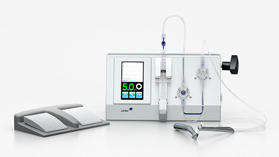
Fig. 1. Jeder-System: pump with foot pedal, drill, and con-
implant are inserted
necting tubing set.
� The whole procedure is constantly monitored by
continuous measurement of the pressure and volumeof the inserted fluid on the display of the Jeder-pump.
retraction. Then, the primary drill is taken until
The Jeder-pump features a built-in security mechanism
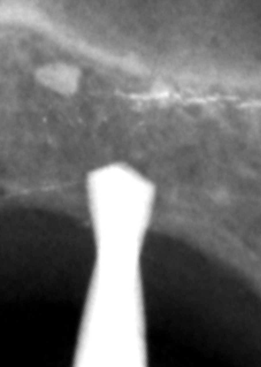
1-2 mm below the sinus floor. To verify depth, an intra-
to prevent introducing excessive pressure and fluid
operative radiograph can be done
volume: As each step on the foot pedal of the Jeder-
Once the primary drilling has reached a sufficient
pump injects only 0.2 mL of saline solution, there is no
depth shortly below the sinus floor, the Jeder-drill is
danger for the Schneiderian membrane after the
plugged into the bore (pressure-sealed) )
perforation of the bone ("high pressure, but very
and high hydraulic pressure (1.5 bar) is built up in the
limited amount of liquid"). Additionally, all data are
pressure chamber of the Jeder-drill using physio-
electronically recorded for documentation purposes.
logical saline solution (NaCl). The centrally placeddrill within the pressure chamber of the Jeder-drill
A combination of Ostim (Heraeus Kulzer, Vienna,
slowly moves through the remaining crestal bone
Austria) and Bio-Oss (Geistlich Biomaterials, Baden-
toward the sinus floor (
Baden, Germany) was used as augmentation material.
� Upon the first minimal perforation of the remaining
In total, 20 Ankylos screw type implants (DENTSPLY-
fluid pushes back the
Friadent, Mannheim, Germany) were placed immedi-
membrane, ensuring that the drill does not perforate
ately after MISFA. Implant diameters were 3.5 mm in
ORAL AND MAXILLOFACIAL SURGERY
296 Jesch et al.
Fig. 4. The ATP-punch, developed by Prof. Dr. WolfgangJesch.
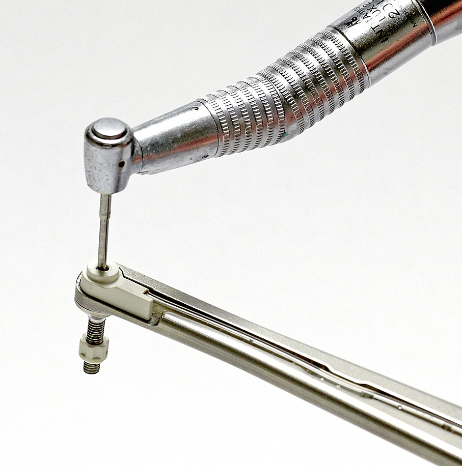
Fig. 2. The Jeder-drill consists of the guide-element and thecentrally placed drill.
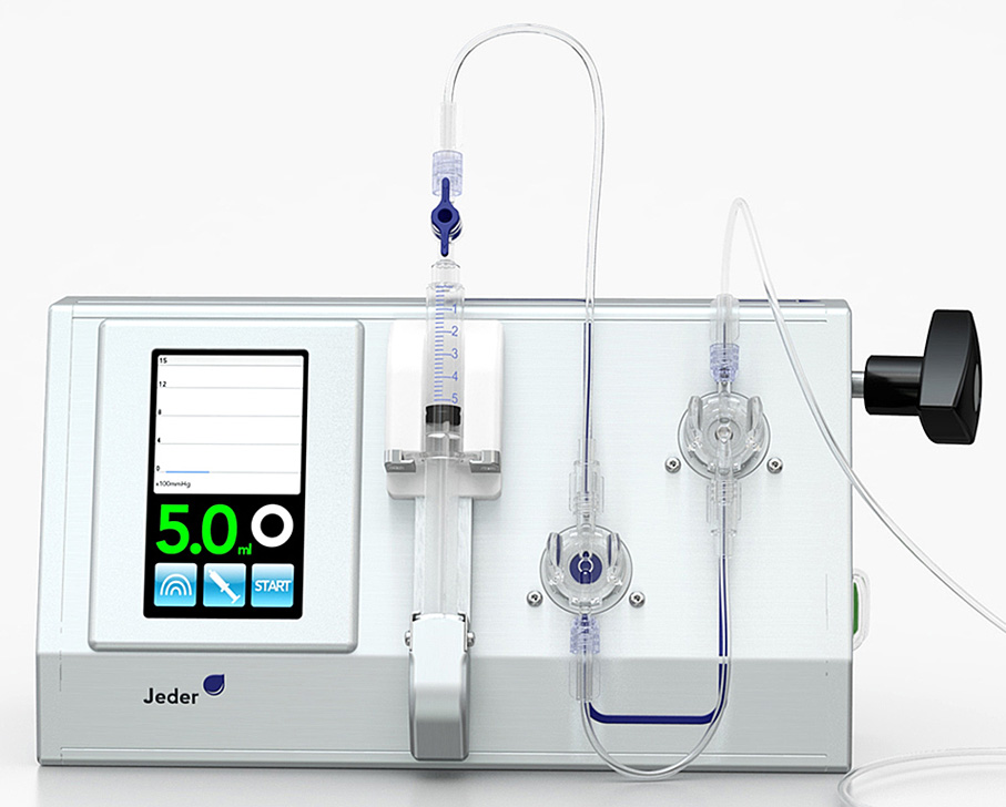
Fig. 3. The Jeder-pump with control display, controlled byfoot pedal.
4 cases and 4.5 mm in 16 cases. Implant length was11 mm in all but 2 cases. One implant was 9.5 mm longwhile another one was 14 mm.
Fig. 5. The depth of the primary drill can be verified bytaking an intra-operative radiograph.
Peri- and postoperative carePrior to surgery local anesthetic infiltration was admin-istered buccally and palatally (articaine hydrochloride 4%
implant load was 258 � 81 days. Prosthetic rehabilitation
with epinephrine 1:100,000 [Septanest with Epinephrine;
included single crowns in 12 cases, implant bridge
Septodont, Niederkassel-Mondorf, Germany]). Postop-
constructions in 6 cases (including 1 horseshoe implant
eratively, patients were prescribed either clindamycin
bridge), and removable partial dentures in 2 patients.
hydrochloride 300 mg (Clindac; Sandoz GmbH, Kundl,Austria) 3 times daily or josamycin 500 mg (Josalid;
Clinical and radiologic follow-up
Sandoz) twice a day, for 1-week period. Following
On day 1 and day 3 after surgery, telephone follow-ups
successful implantation the healing period until full
were conducted. One week postoperatively, every
Volume 116, Number 3
Jesch et al.
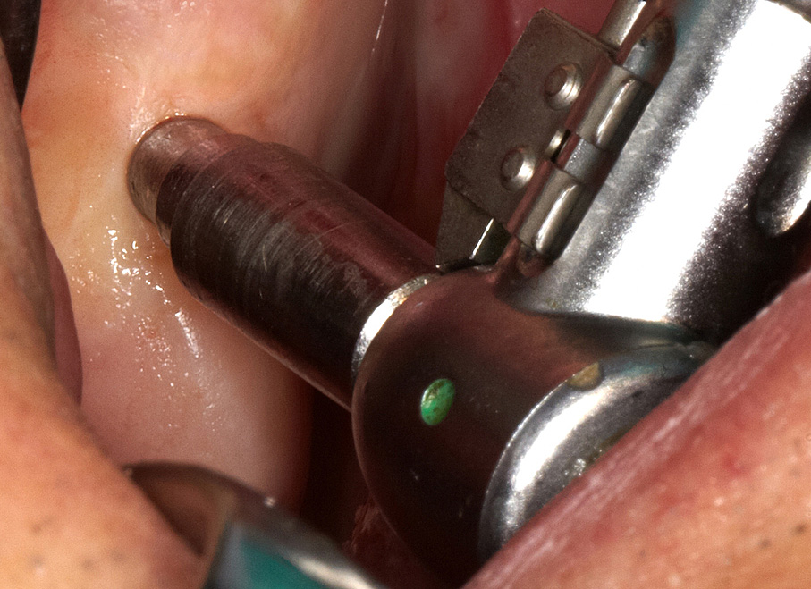
Fig. 6. The sealing element of the Jeder-drill (Jeder GmbH,
Fig. 8. The high pressure pushes the Schneiderian membrane
Vienna, Austria) is pressed against the mucosa.
away from the drill.
absence of clinically detectable implant mobility, (2)absence of pain or any subjective sensation, (3) absenceof recurrent peri-implant infection, and (4) absence ofcontinuous radiolucency around the implant.
Statistical analysisDescriptive statistical analysis including calculation ofmean values and standard deviation (SD) of recordeddata was carried out with Statistica software (release6.0, StatSoft Inc., Tulsa, OK, USA). Data are presentedas means � SD.
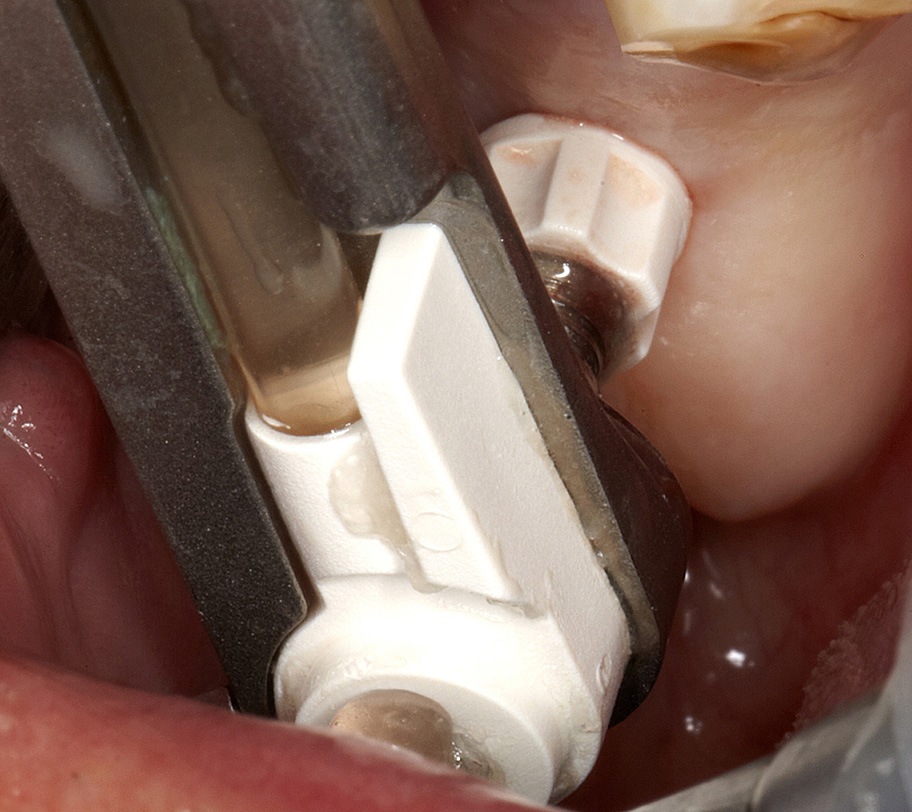
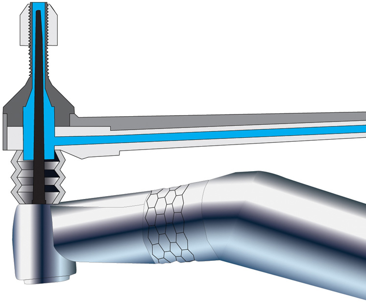
Fig. 7. Hydraulic pressure is built up in the pressure chamber
In total, 20 MISFA were performed in 18 patients. Main
of the Jeder-drill (Jeder GmbH, Vienna, Austria) using
data are summarized in
physiological saline solution.
On the control CT scan performed 4-6 weeks post-
operatively, a small amount of augmentation material
patient completed a questionnaire including the degree
was observed in a maxillary recessus in 1 out of 20 cases.
of overall patient satisfaction e by use of a visual analog
Thus, the membrane perforation rate was 5%. The
scale (VAS) (1 ¼ not satisfied, 10 ¼ very satisfied) e and
patient did not show any clinical symptoms and there
including the days of sick leave. Between 4 and 6 weeks
was no implication for the implant placement. The
after surgery, a clinical examination and a control CT
respective patient is under close observation and at the
scan were performed aiming at detecting possible post-
18-month follow-up there were no clinical symptoms
related to this perforation of the Schneiderian membrane.
membrane perforation) as well as quantification of
The control CT scan also showed that the residual
height gained by the augmentation procedure. All CT
bone height of 4.6 � 1.4 mm could be augmented to
images were evaluated by 2 independent investigators
13.8 � 2.3 mm corresponding to a height gain of
not involved in the surgical procedures.
9.2 � 1.7 mm.
Eighteen months postoperatively (562 � 34 days)
Overall patient satisfaction with the surgical proce-
implant stability was assessed in a clinical and radiologic
dure was evaluated based on a questionnaire using
[periapical radiograph and digital volume tomography
a VAS ranging from 1 to 10 (1 ¼ not satisfied,
(DVT)] examination. The survival criteria proposed by
10 ¼ very satisfied). Average patient satisfaction was
Buser et and Cochran et were followed at the
9.82 � 0.7 points. The mean duration of sick leave was
18-month control. They included the following: (1)
0.19 � 0.5 days.
ORAL AND MAXILLOFACIAL SURGERY
298 Jesch et al.
Fig. 10. The postoperative DVT shows the sinus lift and theimplant in situ.
method for cases with at least 3 mm residual alveolarridge height.
Height gainIt is widely accepted in the literature that crestalmethods currently in use are not predictable andreproducible if elevations >5 mm are needeHowever, in the present study the height gain rangedfrom 6 to 11 mm (mean 9.2 � 1.7 mm). In cadavericstudies it has been demonstrated that in a crestalapproach the Schneiderian membrane can be liftedmore than 10 mm without tearing.Membrane
Fig. 9. The pressure-drop on the display of the Jeder-pump
perforation can occur as soon as elevation forces exceed
(Jeder GmbH, Vienna, Austria) indicates the successfulperforation of the remaining bone.
the load limits of the sinus memAs the Jeder-System uses hydraulic pressure, Pascal's principle ofeven pressure distribution appliesand allows opti-
At the 18-month follow-up, none of the implants
showed any mobility apart from 1 case where the
concluded that the novel method can deliver a height
implant was lost 9 months after surgery. Following
gain that is comparable to the lateral approach.
insertion of a new implant at the same site 3 monthsafter explantation, the new implant is still in place andfunctions well according to the patient's feedback. The
18-month survival rate was therefore 95% (19 of 20
In the literature, perforation rates for indirect sinus
implants). None of the remaining implants showed any
floor augmentations usually vary between 0% and
mobility, and there was no pain, redness, swelling, or
44%.In reality, microscopic tears are, in many
suppuration at any implant site. None of the patients
instances, impossible to diagnose and therefore their
reported clinical symptoms of maxillary sinusitis, and
incidence frequency is often underestimated.Some
also the DVT did not reveal any case of sinusitis.
authorexplicitly state that small perforations(without clinical verification) might not have beendetected, which means that the perforation rates re-
ported in their studies would be too low.
The main finding of our study is that e within the limits
In an endoscopically controlled osteotome sinus
of a prospective open cohort study with 20 cases e
floor augmentation study, the validity of the Valsalva
MISFA using the Jeder-System is a safe and effective
Volume 116, Number 3
Jesch et al.
effectiveness to detect small perforations of the
<4 mm, further studies are needed to verify that the
Schneiderian membrane.In our study, CT scans were
novel method can also yield high survival rates for sites
undertaken before and 4-6 weeks after MISFA in all
with pre-operative bone height of <4 mm.
patients. Additionally, all CT images were evaluated by2 independent investigators not involved in the surgical
procedure. In one patient a small amount of augmen-
All recorded side effects were considered slight or
tation material was radiologically visible in the upper
moderate. In particular, there were no signs of infec-
recessus of the maxillary sinus without any clinical
tion, sinusitis or nose bleeding after MISFA, while
symptoms of sinusitis. Therefore, membrane perfora-
one hematoma was observed after local anesthesia. It
tion rate was considered to be 5% (1 of 20 cases). The
could be shown that the novel method significantly
perforation neither affected implant stability nor caused
reduces side effects compared to the considerably
any problems for the patient. At the 18-month follow-
more invasive lateral approach. Thus, sick leave was
up visit, the patient demonstrated no clinical symptoms
very low in the present study at 0.17 � 0.5 days
or radiological signs of sinusitis and the implant loading
compared to 4.5 � 3.2 days after the lateral procedure
was successful. We can confirm the fithat
(unpublished own data). Overall patient satisfaction
small membrane perforations are compatible with
was 9.87 � 0.7 points on a VAS from 1 ¼ not satisfied
clinically healthy postoperative sinus conditions.
to 10 ¼ very satisfied.
In his systematic review of 10 transcrestal sinus lift
Within the limits of a prospective open cohort study
studies, Tanidentified a reported mean implant
with 20 cases, our data demonstrate the safety and
success rate of 92.8%. This is almost the same as for
medical efficacy of MISFA using the novel method. A
implants placed in the posterior maxilla without graft-
height gain of 9.2 � 1.7 mm could be achieved. In only
ing, i.e., 95.9%-Watzerightly remarks
one case a membrane perforation occurred without any
that the residual local bone height of 6-7 mm usually
clinical consequences. The 18-month survival rate was
recommended for transcrestal sinus floor elevation
95%. Side effects were acceptable and remarkably
means that implants of appropriate length should be
lower compared with the lateral approach. Sick leave
firmly seated in the host bone. Therefore, at least in the
occurrence was very low and overall patient satisfaction
first few years, Watzek considers it difficult to say
very high. To further evaluate the Jeder-System
whether implant stability was achieved because of
a broader clinical study (w100 cases) with international
transcrestal sinus floor elevation or on account of the
study centers is planned in the near future.
bone volume-related short osseointegration segmentsufficient for short implants.
For a residual bone height below the recommended
range, the story is different. Without quantifying the
effect, confirms the finding of Rosenthat the
failure rate of the implants increased with and was
correlated to reduced residual bone height. In a multi-
center retrospective study, Rosen reported a survival
rate of 96%, when the residual bone height was 5 mm
or more, but the survival rate decreased to 85.7%, when
the residual bone height was 4 mm or less. Watzek
5. Rodrigues GN, Katayama AY, Cardoso RF. Subantral Augmen-
rightly remarks that at a residual bone height of
tation Utilizing the Zimmer Sinus Lift Balloon Technique. 2010.
<4 mm, implant survival has rarely been reported. In
his own recent study with the gel-pressure technique,
Accessed March 3, 2013.
Watzereports an implant survival rate 1 year after
implant placement of 88.5% at sites with a pre-opera-
tive bone height of at least 4 mm (n ¼ 26). This
decreases to a 1 year survival rate of 50.0% for sites
with a pre-operative bone height of <4 mm (n ¼ 14).
In our study, the survival rate 18 months after implant
placement was 94.1% at sites with a pre-operative bone
height of at least 4 mm (n ¼ 17) and 100% for sites with
a pre-operative bone height of <4 mm (n ¼ 3). Due to
the small number of cases with residual bone height of
ORAL AND MAXILLOFACIAL SURGERY
300 Jesch et al.
Reprint requests:
Emanuel Bruckmoser, MD, DMD
Department of Oral and Maxillofacial Surgery
ABM University Health Board
Morriston Hospital
Swansea SA6 6NL, Wales, UK
Source: http://www.sinusafe.co.il/image/users/270045/ftp/my_files/Jeder_article_OOOO_Sep_2013_DB-1.pdf?id=15750355
DESIGN GUIDE The Royal Parks Design guide, technical specifi cations D e s i g n g u i D eMaintaining the historic landscape the royal parks Context the eight royal parks comprise Bushy nash and Charles Bridgeman. park, the green park, greenwich the quality of the landscape design is park, hyde park, kensington gardens,
ABRACADAVER MAGAZINE, Página 2 Número 1, Octubre de 2003. Editorial Has estado bajo el habla de personas que son menos que tú y que se creen con el derecho de decir y criticar todo lo que en tu vida haces. Sientes que muchas otras apenas tienen la cultura medianamente aceptable y aún así, te tratan como si no pertenecieras a este mundo. Que importa que, a los ojos de la gente, tengas un problema… En realidad estás aquí para hacerles recordar que son ellos los del problema…Lo único que nos mueve a escribir en Abracadaver es esa sensación de que algo anda mal y de que


