Kamagra gibt es auch als Kautabletten, die sich schneller auflösen als normale Pillen. Manche Patienten empfinden das als angenehmer. Wer sich informieren will, findet Hinweise unter kamagra kautabletten.
Apoptosis in vascular disease

Eur J Vasc Endovasc Surg 22, 389–395 (2001)
doi:10.1053/ejvs.2001.1492, available online at http://www.idealibrary.com on
Apoptosis in Vascular Disease
T. C. F. Sykes∗1,2, A. G. Morris1, A. W. Bradbury2 and D. Mosquera2
1Biological Sciences, University of Warwick, Coventry CV4 7AL, 2Department of Vascular Surgery,
Birmingham Heartlands Hospital, Birmingham B9 5SS, U.K.
process may offer novel avenues for therapeutic inter-
Apoptosis, or programmed cell death, was first de-
scribed by Kerr et in 1972 as a specific type of cell
death morphologically distinct from necrosis. Apo-
Molecular Aspects of Apoptosis
ptotic cells undergo shrinkage with nuclear con-
densation and the formation of membrane-bound Since the original morphological description of apo-
vesicles called apoptotic bodies. These are phago- ptosis many molecular mechanisms that characterise
cytosed by adjacent cells and macrophages without the apoptotic pathway have been identified. Apoptosis
inducing an inflammatory response. In contrast, nec- is controlled by a well-ordered cascade of cellular
rotic cells sustain membrane damage leading to the events that may be divided into four stages (initiation,
release of pro-inflammatory intracellular contents. control/execution, structural/morphological al-
Apoptosis although recognised to be fundamentally teration and phagocytic recognition) (Fig. 1). Initiation
important in development also occurs in normal of apoptosis may occur following the binding of death
physiological circumstances. Apoptotic vascular promoting factors (e.g. Tumour Necrosis Factor (TNF)
smooth muscle cells (VSMCs) have been identified and Fas Ligand) to cell surface TNF and Fas death
during the physiological regression and closure of receptors or by a withdrawal of survival factors (e.g.
the human ductus arteriosus before Similarly, reduced insulin-like growth factor, loss of cell–cell
apoptotic VSMCs and endothelial cells are present in and cell–matrix contact) acting via the mitochondria.
umbilical veins and arteries following the significant Alternatively apoptosis may be initiated by ionising
haemodynamic changes that occur during Such radiation or chemotherapeutic agents acting directly
apoptosis appears to be triggered by a reduction in on DNA.
blood flow. Further support comes from experimental
The control and execution stage of apoptosis is
studies using immature rabbits which demonstrate dependent on the sequential activation of a group of
that reducing flow in the common carotid artery, by cysteine proteases referred to as caspases. Activation
ligating the external carotid, results in both VSMC and ofcaspase-8occursfollowingbindingofthecellsurface
endothelial cell The normal blood vessel death-receptors (TNF and Caspase-9 is activated
wall is characterised by a low endothelial cell turnover by cytochrome C released from mitochondria stim-
with a variety of mechanisms affording protection ulated by the withdrawal of survival factors. Both
against Indeed, recent data indicate that caspase-8 and 9 can activate the final executioner,
apoptosis may be important in the pathophysiology caspase-3, which in turn causes the irreversible frag-
of vascular disease. A better understanding of this mentation and degradation of DNA.
Control of the execution phase is achieved by anti-
∗ Please address all correspondence to: T. C. F. Sykes, Department
apoptotic proteins belonging to the Bcl-2 family (Bcl-
of Vascular Surgery, Research Institute, Lincoln House, Birmingham
Heartlands Hospital, Birmingham B9 5SS, U.K.
2, Bcl-XL) inhibiting pro-apoptotic proteins (Bax and
1078–5884/01/110389+07 $35.00/0 2001 Harcourt Publishers Ltd.
T. C. F. Sykes et al.
Fig. 2. A schematic summary of pro-apoptotic and survival factors
typically affecting the endothelial cell. A loss of survival factors will
also precipitate apoptosis. ROS (reactive oxygen species), VEGF
(Vascular endothelial cell growth factor).
apoptosis in tissue sections. The major drawback of
Fig 1. A schematic diagram illustrating four functional stages of
the TUNEL technique is that nuclei can be stained
apoptosis and the three major pathways through which the cascade
non-specifically due to differences in cell fixation and
of cellular events can proceed. The three pivotal caspases (8, 9
also non-nuclear structures in atherosclerotic plaques
and 3) are shown with caspase-3, the final executioner, causing
irreversible DNA damage and loss of cell viability. Control is
may be stained. It is recommended that this technique
achieved with the regulatory pro and anti-apoptotic proteins (Bax/
be used in conjunction with an additional mor-
Bid & Bcl2 respectively). TNF (Tumour necrosis factor/receptor),
Fas L (Fas Ligand), Fas (Fas receptor), DD (TNF death domain
protein), FADD (Fas-associated death domain), IGF (Insulin-like
Additional methods, suitable for in vitro use only,
growth factor) and PS (Phosphatidylserine).
phatidylserine (PS) residues, that occurs in early apo-
Bid) that facilitate the release of cytochrome C from the ptosis, with a fluorescein (FITC) conjugate of Annexin-
mitochondria. The relative amounts of these proteins V, a protein that has a high natural affinity for PS.
determines whether the cell undergoes apoptosis or This method allows quantitative assessment of apo-
not. Structural and morphological changes to the cell ptotic cells either by fluorescence microscopy or flow
occur following the internucleosomal fragmentation cytometry.
of DNA leading to an irreversible loss of cell viability.
Early apoptotic cells are characterised by ex-
ternalisation of phosphatidylserine (PS) residues,
Pro-apoptotic Factors Implicated in Vascular Cell
which are normally situated on the inner leaflet of the
plasma membrane. This feature facilitates the specific
recognition of apoptotic cells by macrophages enabling In vitro studies have shown that extracellular agents,
their elimination by phagocytosis.
including well characterised cardiovascular risk fac-
The efficient removal of apoptotic cells can make tors and inflammatory mediators induce apoptosis of
their identification difficult. Historically the detection endothelial cells and vascular SMCs. (Fig. 2).
and quantification of apoptotic cells has relied on
morphological assessment by electron microscopy or
light Newer techniques rely on the de-
tection of fragmented DNA by terminal transferase
Oxidised Low Density Lipoprotein (OxLDL)
mediated dUTP-biotin nick end labelling (TUNEL) or
in situ nick translation Both methods have Elevated LDL is a significant risk factor for coronary
become the standard technique for the detection of artery disease. Specifically, its oxidised form (OxLDL)
Eur J Vasc Endovasc Surg Vol 22, November 2001
Apoptosis in Vascular Disease
is considered to play a key role in the development
of premature atherosclerosis through the formation of
foam cells and fatty OxLDL is known to be
cytotoxic to cultured endothelial cells and has also
been shown to induce apoptosis, in bovine aortic
endothelial cells and human umbilical vein endothelial
cells OxLDL-induced apoptosis is in-
hibited by the addition of the antioxidant vitamins C
and and is therefore believed to act through the
generation of reactive oxygen species. Cultured vas-
cular smooth muscle cells also undergo apoptosis
when exposed to oxLDL and
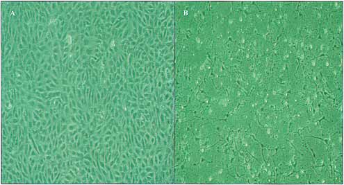
Fig. 3. Phase contrast microscopy of cultured human umbilical
venous endothelial cells (HUVECs). A: control cells with a char-
acteristic cobblestone morphology, B: After 4 h incubation with
homocysteine and adenosine (0.5 mM) showing contracted apoptotic
Hyperglycaemia/advanced glycation end products
Cytokines and inflammatory mediators
Endothelial cells cultured with high concentrations of The inflammatory cytokines TNF!, IL-1" and IFN-
glucose (30 mmol/L) for 48 h show increased apoptosis # have all been identified in human atherosclerotic
compared with those incubated in low concentrations TNF! causes endothelial cell
of glucose (5 High glucose also appears to through the activation of caspase-3 and may be in-
induce apoptosis through the generation of hydrogen hibited by specific inhibitors of this In-
peroxide and can be inhibited by antioxidants such terestingly, TNF! can also activate a survival pathway
as ascorbic Similarly high concentrations of that requires protein Inhibitors of RNA
advanced glycation end products, the products of non- transcription or protein synthesis can therefore aug-
enzymatic glycation of proteins in the circulation, can ment TNF!-induced apoptosis of endothelial cells. In
induce apoptosis of endothelial cells after 48 In- contrast TNF!, does not cause apoptosis of cultured
terestingly, the lipid-lowering drug pravastatin can human smooth muscle cells on its own, but does so
prevent hyperglycaemia induced which when combined with IL-1" and IFN-Lipo-
contrasts with the proapoptotic effects of the lipophilic polysaccharide also induces human endothelial cell
statins (atorvastatin, simvastatin and lovastatin) on apoptosis in vitro that can be prevented by anti-
vascular smooth muscle
This effect also occurs in vivo, but unlike
the in vitro setting is dependent on TNF!
Angiotensin II, although promoting growth of VSMCs,
induces endothelial apoptosis in vitro in a dose de-
Hyperhomocysteinaemia is, an independent risk factor
for cardiovascular disease, and, is known to cause
Reactive oxygen species
endothelial We have recently shown
that homocysteine in combination with adenosine, an Elevated reactive oxygen species (ROS) are an early
important substrate in homocysteine metabolism, can hallmark of In vitro evidence indicates
induce apoptosis of cultured HUVECs and human that ROS, the by-product of aerobic metabolism, in-
duce endothelial and smooth muscle cell
long saphenous vein endothelial cells after as little as
4 h (Fig. 3). These effects can be prevented ROS may be produced in the vessel wall by macro-
by vitamin B6, B12 and cofactors in homo- phages within the atherosclerotic plaque or endo-
cysteine metabolism and currently used as homo- genously by human endothelial and vascular smooth
cysteine-lowering agents in clinical trials. The latter muscle cells. The proatherosclerotic and proapoptotic
finding may represent an important mechanism by factors oxLDL, TNF!, glucose and angiotensin II, all
which vitamin supplementation can reduce vascular induce endogenous ROS whose effects can be pre-
injury associated with hyperhomocysteinaemia.
Eur J Vasc Endovasc Surg Vol 22, November 2001
T. C. F. Sykes et al.
Survival Factors Implicated in Preventing
Vascular Cell Apoptosis
The survival of endothelial cells and vascular smooth
Shear stress and nitric oxide
muscle cells is dependent on contact with adjacent
cells and the extracellular matrix (Fig. 2). This is me-
Cultured endothelial cells (HUVECs) undergo a basal diated by cellular adhesion molecules such as the
level of apoptosis in the absence of flow, which can be integrins, which also function as signalling molecules.
inhibited by mimicking flow conditions in a perfusion Apoptosis occurs when these contacts are Under
chamberLaminar flow generates shear stress at the normal circumstances the extracellular matrix gen-
endothelial cell surface that can prevent apoptosis erates survival signals that either suppress apoptotic
induced by different stimuli including TNF!, oxLDL pathways or lead to an increase in the activity of
and ROS. This inhibition is mediated by shear stress- anti-apoptotic pathways such as the Bcl-2 family of
induced release of nitric oxide (NO) that subsequently
inactivates In the normal vasculature the
shear stress associated with laminar flow causes a
continuous production of NO by endothelial cells,
providing protection from injury and apoptosis. How-
Apoptosis and Vascular Disease
ever atherosclerotic plaque-prone areas are typically
sites of turbulent blood flow and low shear stress, and
are associated with increased cell which is
most probably secondary to increased
Early studies in cholesterol-fed swine have shown
cell death to be a major component of atherosclerotic
plaque In situ techniques (TUNEL)
have since confirmed the presence of apoptotic smooth
muscle cells, T-lymphocytes, and macrophages in
Growth factors
human atherosclerotic More recently
Tricot et have examined carotid endarterectomy
Cultured endothelial cells deprived of growth factor specimens and shown that apoptotic luminal endo-
undergo Addition of vascular endothelial thelial cells occur with a greater prevalence in the
growth factor (VEGF) can inhibit apoptosis induced post-stenotic area, a region of low flow and low shear
by TNF!ionising and disruption of the stress.
extracellular Angiopoietin-1 can also prevent
Apoptotic cell death within plaques, as determined
apoptosis in growth factor deprived endothelial by the TUNEL technique, ranges from <2% to 30% and
Basic fibroblast growth factor (FGF-2) prevents lipo- is related to the stage of the atherosclerotic
popolysaccharide induced endothelial cell apoptosis in Very little apoptosis occurs in intimal thickening
vivo and serum and growth factor deprivation induced and fatty streaks with the majority of apoptotic cells
apoptosis in In cultured human vascular occurring in advanced atherosclerotic plaques within
smooth muscle cells several growth factors (insulin- regions of macrophage
like growth factor, platelet derived growth factor, basic
Although the above evidence indicates that apo-
fibroblast growth factor, and transforming growth fac- ptosis does occur within human atherosclerotic
tor) inhibit apoptosis especially under low serum con- plaques, other authors have used electron microscopy
to show that despite >10% TUNEL-positive nuclei, the
vast majority of injured and disintegrating cells within
plaques display typical features of cells undergoing
Although the relative importance of cell death by
apoptosis versus necrosis is unknown we can speculate
Oestrogen is an established atheroprotective hormone, that apoptosis may allow plaque stabilisation. Apo-
which is known to produce beneficial changes in lipid ptotic removal of T-cells and macrophages, which
profile and the regulation of vascular tone. Oestradiol commonly infiltrate the shoulder region of the fibrous
also maintains endothelial integrity by inhibiting endo- cap, would reduce matrix metalloproteinase (MMP)
thelial apoptosis induced byTNF!Antioxidant vit- synthesis and extracellular matrix breakdown without
amins C and E, especially in combination, prevent an accompanying inflammatory reaction. Conversely
apoptosis induced by oxLDL, TNF!, and
death of vascular smooth muscle cells in plaques,
Eur J Vasc Endovasc Surg Vol 22, November 2001
Apoptosis in Vascular Disease
either through apoptosis or necrosis, may lead to a
weakening of the fibrous cap as a consequence of
reduced collagen and extracellular matrix synthesis Medial SMC loss prior to the development of occlusive
following the loss of smooth muscle cells.
and intimal proliferative lesions is characteristically
Apoptosis of luminal endothelial in athero- found in transplant arteriopathy. Experimental evi-
sclerotic plaques may initiate plaque erosion with dence using rat arterial allografts suggest that this
the subsequent promotion of platelet aggregation and SMC loss is due to Endothelial damage,
thrombosis. Apoptotic vascular cells themselves may also seen in transplant arteriopathy, appears to be due
also lead to increased plaque thrombogenicity. Both to a Fas-based apoptotic
apoptotic smooth muscle cells and endothelial cells
expose phosphatidylserine residues on their surface
in early apoptosis, which in the presence of factor V
and VII, can act as a substrate for the generation
of Additionally apoptotic monocytic cells Apoptoticcellsarequicklyeliminatedbyneighbouring
have enhanced tissue factor Mallat et phagocytic cells making in vivo identification difficult.
have also identified increased tissue factor expression Our current understanding of apoptosis in vascular
around apoptotic cells within the central necrotic core disease therefore depends almost entirely upon in vitro
of plaques, indicating that tissue factor is shed from and in situ data. However, recent techniques, which
apoptotic cells via apoptotic microparticles.
capture shed microparticles released by apoptotic cells,
have been used to show increased apoptosis in acute
Induction of apoptosis
Apoptosis of smooth muscle cells will occur following
balloon injury in animal models. An initial apoptotic Atherosclerotic plaques in cholesterol-fed rabbits re-
response after 30 is followed by persistent apo- gress when neointimal cell apoptosis is induced by
ptosis after two weeks, the latter associated with the inhibiting the antiapoptotic protein Plaque re-
regulation of intimal Studies of human gression also occurs following administration of L-
restenotic plaques indicate there may be an increased arginine to induce macrophage apoptosis via NO re-
apoptotic rate, but there is no consensus. Isner et Interestingly, Schaub et found that when
found an increased apoptotic rate in human restenotic vascular smooth muscle cell apoptosis is induced,
plaques, whereas Bauriedel et noted reduced levels through the overexpression of death domain proteins,
of apoptotic cells in restenotic plaques compared with macrophages are recruited and neointimal progression
primary plaques.
rather than regression occurs.
Various attempts to induce apoptosis have been
tried in animal models of neointima formation and
restenosis following angioplasty. Transfection of sui-
cide genes into smooth muscle cells or direct delivery
of pro-apoptotic ligands (Fas-ligand) into the vessel
wall induce smooth muscle cell apoptosis and a sig-
nificant reduction in neointima formation in rabbit
Reduced smooth muscle cell density within the elastic arteries following Adopting this ap-
media of human aneurysmal wall is associated with proach in the human clinical setting may however,
increased smooth muscle cell Further risk the development of aneurysms.
evidence has shown that infiltrating T cells express
cytotoxic mediators such as cytokines, perforin and
Fas/Fas ligand, which are capable of inducing apo-
ptosis. It is hypothesised that these might contribute
Inhibition of apoptosis
to the elimination of smooth muscle cells, a source of
collagen and elastin, thereby impairing repair and Currently there is no information on the effect of
maintenance of the arterial extracellular matrix tending inhibiting apoptosis on the progression or com-
to favour aneurysmal wall expansion.
plications of vascular disease. In vitro and in situ data
Eur J Vasc Endovasc Surg Vol 22, November 2001
T. C. F. Sykes et al.
LDLs induce massive apoptosis of cultured human endothelial
indicate that apoptosis of many cell types occurs in
cells through a calcium-dependent pathway. Prevention by au-
human atherosclerotic plaques, and aneurysmal wall.
rintricarboxylic acid. Arterioscler Thromb Vasc Biol 1997; 17: 331–
Strategies to inhibit apoptosis in these cell types may be
12 Haendeler J, Zeiher AM, Dimmeler S. Vitamin C and E prevent
the best approach to limit plaque erosion, thrombosis,
lipopolysaccharide-induced apoptosis in human endothelial cells
progression and aneurysmal development. Potential
by modulation of Bcl-2 and Bax. Eur J Pharmacol 1996; 317:
therapeutic options include the use of anti-apoptotic
13 Jovinge S, Crisby M, Thyberg J, Nilsson J. DNA fragmentation
agents known to inhibit apoptosis in vitro including
and ultrastructural changes of degenerating cells in ath-
NO, growth factors (VEGF), angiotensin converting
erosclerotic lesions and smooth muscle cells exposed to oxidized
enzyme inhibitors, antioxidants and vitamins (B6, B12
LDL in vitro. Arterioscler Thromb Vasc Biol 1997; 17: 2225–2231.
and folate) as well as controlling the levels of es- 14 Nishio E, Watanabe Y. Oxysterols induced apoptosis in cultured
smooth muscle cells through CPP32 protease activation and bcl-
tablished cardiovascular risk factors (lipids, glucose,
2 protein downregulation. Biochem Biophys Res Commun 1996;
and homocysteine).
15 Baumgartner-Parzer SM, Wagner L, Pettermann M et al.
High-glucose-triggered apoptosis in cultured endothelial cells.
Diabetes 1995; 44: 1323–1327.
16 Ho FM, Liu SH, Liau CS, Huang PJ, Lin-Shiau SY. High glucose-
induced apoptosis in human endothelial cells is mediated by
sequential activations of c-Jun NH(2)-terminal kinase and ca-
spase-3. Circulation 2000; 101: 2618–2624.
Despite the wealth of in situ and in vitro data dem- 17 Min C, Kang E, Yu SH, Shinn SH, Kim YS. Advanced glycation
onstrating that apoptosis not only occurs in vascular
end products induce apoptosis and procoagulant activity in
disease, but can be induced by established cardio-
cultured human umbilical vein endothelial cells. Diabetes Res
Clin Pract 1999; 46: 197–202.
vascular risk factors, there is little evidence in humans 18 Moneley D, Hong C, Condron C et al. Pravastatin prevents
to indicate whether apoptosis is beneficial or harmful.
hyperglycaemia-induced human vascular endothelial cell apo-
Direct modulation of apoptosis may provide thera-
ptosis. Br J Surg 2001; 88: 748.
19 Guijarro C, Blanco-Colio LM, Massy ZA et al. Lipophilic
peutic avenues to alter the pathophysiology of vascular
statins induce apoptosis of human vascular smooth muscle cells.
disease. However, to date, despite successes in some
Kidney Int Suppl 1999; 71: S88–S91.
animal models, their applicability to human vascular 20 Refsum H, Ueland PM, Nygard O, Vollset SE. Homocysteine
and cardiovascular disease. Annu Rev Med 1998; 49: 31–62.
disease remains unknown.
21 Sykes TCF, Molostvov G, Morris A, Mosquera D. Homo-
cysteine induces apoptosis in human vascular endothelium. Br
J Surg 2001; 88: 748.
22 Sykes TCF, Molostvov G, Morris A, Mosquera D. Homo-
cysteine-induced apoptosis is inhibited by vitamin B6, B12, and
folate. Br J Surg 2001; 88: 4.
23 Libby P, Sukhova G, Lee RT, Galis ZS. Cytokines regulate
1 Kerr JF, Wyllie AH, Currie AR. Apoptosis: a basic biological
vascular functions related to stability of the atherosclerotic
phenomenon with wide-ranging implications in tissue kinetics.
plaque. J Cardiovasc Pharmacol 1995; 25: S9–S12.
Br J Cancer 1972; 26: 239–257.
24 Robaye B, Mosselmans R, Fiers W, Dumont JE, Galand P.
2 Slomp J, Gittenberger-de Groot AC, Glukhova MA et al.
Tumor necrosis factor induces apoptosis (programmed cell
Differentiation, dedifferentiation, and apoptosis of smooth
death) in normal endothelial cells in vitro. Am J Pathol 1991; 138:
muscle cells during the development of the human ductus
arteriosus. Arterioscler Thromb Vasc Biol 1997; 17: 1003–1009.
25 Dimmeler S, Haendeler J, Nehls M, Zeiher AM. Suppression
3 Kim HS, Hwang KK, Seo JW et al. Apoptosis and regulation
of apoptosis by nitric oxide via inhibition of interleukin-1 beta-
of Bax and Bcl-X proteins during human neonatal vascular
converting enzyme (ICE)-like and cysteine protease protein
remodeling. Arterioscler Thromb Vasc Biol 2000; 20: 957–963.
(CPP)-32-like proteases. J Exp Med 1997; 185: 601–607.
4 Cho A, Mitchell L, Koopmans D, Langille BL. Effects of
26 Karsan A, Yee E, Harlan JM. Endothelial cell death induced
changes in blood flow rate on cell death and cell proliferation
by tumor necrosis factor-alpha is inhibited by the Bcl-2 family
in carotid arteries of immature rabbits. Circ Res 1997; 81: 328–337.
member, A1. J Biol Chem 1996; 271: 27201–27204.
5 Kockx MM, Knaapen MW. The role of apoptosis in vascular
27 Geng YJ, Libby P. Evidence for apoptosis in advanced human
disease. J Pathol 2000; 190: 267–280.
atheroma. Colocalization with interleukin-1 beta-converting en-
6 Ashkenazi A, Dixit VM. Death receptors: signaling and mod-
zyme. Am J Pathol 1995; 147: 251–266.
ulation. Science 1998; 281: 1305–1308.
7 Gavrieli Y, Sherman Y, Ben Sasson SA. Identification of pro-
28 Haimovitz-Friedman A, Cordon-Cardo C, Bayoumy S et al.
grammed cell death in situ via specific labeling of nuclear DNA
Lipopolysaccharide induces disseminated endothelial apoptosis
fragmentation. J Cell Biol 1992; 119: 493–501.
requiring ceramide generation. J Exp Med 1997; 186: 1831–1841.
8 Witztum JL, Steinberg D. Role of oxidized low density lipo-
29 Dimmeler S, Rippmann V, Weiland U, Haendeler J, Zeiher
protein in atherogenesis. J Clin Invest 1991; 88 : 1785–1792.
AM. Angiotensin II induces apoptosis of human endothelial
9 Lizard G, Deckert V, Dubrez L et al. Induction of apoptosis in
cells. Protective effect of nitric oxide. Circ Res 1997; 81: 970–976.
endothelial cells treated with cholesterol oxides. Am J Pathol
30 Dimmeler S, Zeiher AM. Reactive oxygen species and vascular
1996; 148: 1625–1638.
cell apoptosis in response to angiotensin II and pro-ath-
10 Dimmeler S, Haendeler J, Galle J, Zeiher AM. Oxidized low-
erosclerotic factors. Regul Pept 2000; 90: 19–25.
density lipoprotein induces apoptosis of human endothelial cells
31 Li PF, Maasch C, Haller H, Dietz R, von Harsdorf R.
by activation of CPP32-like proteases. A mechanistic clue to the
Requirement for protein kinase C in reactive oxygen species-
‘‘response to injury'' hypothesis. Circulation 1997; 95: 1760–1763.
induced apoptosis of vascular smooth muscle cells. Circulation
11 Escargueil-Blanc I, Meilhac O, Pieraggi MT et al. Oxidized
1999; 100: 967–973.
Eur J Vasc Endovasc Surg Vol 22, November 2001
Apoptosis in Vascular Disease
32 Kaiser D, Freyberg MA, Friedl P. Lack of hemodynamic forces
replication and apoptosis in atherosclerotic plaques of cho-
triggers apoptosis in vascular endothelial cells. Biochem Biophys
lesterol-fed rabbits. Atherosclerosis 1996; 120: 115–124.
Res Commun 1997; 231: 586–590.
54 Han DK, Haudenschild CC, Hong MK et al. Evidence for
33 Dimmeler S, Haendeler J, Rippmann V, Nehls M, Zeiher AM.
apoptosis in human atherogenesis and in a rat vascular injury
Shear stress inhibits apoptosis of human endothelial cells. FEBS
model. Am J Pathol 1995; 147: 267–277.
Lett 1996; 399: 71–74.
55 Mallat Z, Ohan J, Leseche G, Tedgui A. Colocalization of
34 Caplan BA, Schwartz CJ. Increased endothelial cell turnover
CPP-32 with apoptotic cells in human atherosclerotic plaques.
in areas of in vivo Evans Blue uptake in the pig aorta. Athero-
Circulation 1997; 96: 424–428.
sclerosis 1973; 17: 401–417.
56 Crisby M, Kallin B, Thyberg J et al. Cell death in human
35 Dimmeler S, Hermann C, Zeiher AM. Apoptosis of endothelial
atherosclerotic plaques involves both oncosis and apoptosis.
cells. Contribution to the pathophysiology of atherosclerosis?
Atherosclerosis 1997; 130: 17–27.
Eur Cytokine Netw 1998; 9: 697–698.
57 Bobryshev YV, Babaev VR, Lord RS, Watanabe T. Cell death
36 Karsan A, Yee E, Poirier GG et al. Fibroblast growth factor-
in atheromatous plaque of the carotid artery occurs through
2 inhibits endothelial cell apoptosis by Bcl-2-dependent and
necrosis rather than apoptosis. In Vivo 1997; 11: 441–452.
independent mechanisms. Am J Pathol 1997; 151: 1775–1784.
58 Ball RY, Stowers EC, Burton JH et al. Evidence that the
37 Karsan A. Tumour necrosis factor and endothelial cell death.
death of macrophage foam cells contributes to the lipid core of
Trends Cardiovasc Med 1998; 8: 19–24.
atheroma. Atherosclerosis 1995; 114: 45–54.
38 Katoh O, Tauchi H, Kawaishi K, Kimura A, Satow Y. Ex-
59 Flynn PD, Byrne CD, Baglin TP, Weissberg PL, Bennett MR.
pression of the vascular endothelial growth factor (VEGF) re-
Thrombin generation by apoptotic vascular smooth muscle cells.
ceptor gene, KDR, in hematopoietic cells and inhibitory effect
Blood 1997; 89: 4378–4384.
of VEGF on apoptotic cell death caused by ionizing radiation.
60 Aupeix K, Toti F, Satta N, Bischoff P, Freyssinet JM. Oyxsterols
Cancer Res 1995; 55: 5687–5692.
induce membrane procoagulant activity in monocytic THP-1
39 Watanabe Y, Dvorak HF. Vascular permeability factor/vascular
cells. Biochem J 1996; 314 (Pt 3): 1027–1033.
endothelial growth factor inhibits anchorage-disruption-induced
61 Mallat Z, Hugel B, Ohan J et al. Shed membrane microparticles
apoptosis in microvessel endothelial cells by inducing scaffold
with procoagulant potential in human atherosclerotic plaques:
formation. Exp Cell Res 1997; 233: 340–349.
a role for apoptosis in plaque thrombogenicity. Circulation 1999;
40 Kwak HJ, So JN, Lee SJ, Kim I, Koh GY. Angiopoietin-1 is an
apoptosis survival factor for endothelial cells. FEBS Lett 1999;
62 Perlman H, Maillard L, Krasinski K, Walsh K. Evidence for
the rapid onset of apoptosis in medial smooth muscle cells after
41 McCarthy NJ, Bennett MR. The regulation of vascular smooth
balloon injury [see comments]. Circulation 1997; 95: 981–987.
muscle cell apoptosis. Cardiovasc Res 2000; 45: 747–755.
63 Bochaton-Piallat ML, Gabbiani F, Redard M, Desmouliere
42 Bai H, Pollman MJ, Inishi Y, Gibbons GH. Regulation of
A, Gabbiani G. Apoptosis participates in cellularity regulation
vascular smooth muscle cell apoptosis. Modulation of bad by a
during rat aortic intimal thickening. Am J Pathol 1995; 146:
phosphatidylinositol 3-kinase-dependent pathway. Circ Res 1999;
64 Bauriedel G, Schluckebier S, Hutter R et al. Apoptosis in
43 Pollman MJ, Naumovski L, Gibbons GH. Vascular cell apo-
restenosis versus stable-angina atherosclerosis: implications for
ptosis: cell type-specific modulation by transforming growth
the pathogenesis of restenosis. Arterioscler Thromb Vasc Biol 1998;
factor-beta1 in endothelial cells versus smooth muscle cells.
Circulation 1999; 99: 2019–2026.
65 Holmes DR, Lopez-Candales A, Liao S, Thompson RW.
44 Fox JC, Shanley JR. Antisense inhibition of basic fibroblast
Smooth muscle cell apoptosis and p53 expression in human
growth factor induces apoptosis in vascular smooth muscle cells.
abdominal aortic aneurysms. Ann NY Acad Sci 1996; 800: 286–
J Biol Chem 1996; 271: 12578–12584.
45 Spyridopoulos I, Sullivan AB, Kearney M, Isner JM, Losordo
66 Lopez-Candales A, Holmes DR, Liao S et al. Decreased vascular
DW. Estrogen-receptor-mediated inhibition of human endo-
smooth muscle cell density in medial degeneration of human
thelial cell apoptosis. Estradiol as a survival factor. Circulation
abdominal aortic aneurysms. Am J Pathol 1997; 150: 993–1007.
1997; 95: 1505–1514.
67 Thompson RW, Liao S, Curci JA. Vascular smooth muscle cell
46 Re F, Zanetti A, Sironi M, Polentarutti N et al. Inhibition
apoptosis in abdominal aortic aneurysms. Coron Artery Dis 1997;
of anchorage-dependent cell spreading triggers apoptosis in
cultured human endothelial cells. J Cell Biol 1994; 127: 537–546.
68 Hirsch GM, Kearsey J, Burt T, Karnovsky MJ, Lee T. Medial
47 Stromblad S, Cheresh DA. Integrins, angiogenesis and vascular
smooth muscle cell loss in arterial allografts occurs by cytolytic
cell survival. Chem Biol 1996; 3: 881–885.
cell induced apoptosis. Eur J Cardiothorac Surg 1998; 14: 89–96.
48 Thomas WA, Reiner JM, Florentin FA, Lee KT, Lee WM.
69 Dong C, Redenbach D, Wood S et al. The pathogenesis of
cardiac allograft vasculopathy. Curr Opin Cardiol 1996; 11: 183–
Population dynamics of arterial smooth muscle cells. V. Cell
proliferation and cell death during initial 3 months in athero-
70 Mallat Z, Benamer H, Hugel B et al. Elevated levels of shed
sclerotic lesions induced in swine by hypercholesterolemic diet
membrane microparticles with procoagulant potential in the
and intimal trauma. Exp Mol Pathol 1976; 24: 360–374.
peripheral circulating blood of patients with acute coronary
49 Hegyi L, Skepper JN, Cary NR, Mitchinson MJ. Foam cell
syndromes. Circulation 2000; 101: 841–843.
apoptosis and the development of the lipid core of human
71 Pollman MJ, Hall JL, Mann MJ, Zhang L, Gibbons GH.
atherosclerosis. J Pathol 1996; 180: 423–429.
Inhibition of neointimal cell bcl-x expression induces apoptosis
50 Bjorkerud S, Bjorkerud B. Apoptosis is abundant in human
and regression of vascular disease. Nat Med 1998; 4: 222–227.
atherosclerotic lesions, especially in inflammatory cells (macro-
72 Wang BY, Ho HK, Lin PS et al. Regression of atherosclerosis:
phages and T cells), and may contribute to the accumulation of
role of nitric oxide and apoptosis. Circulation 1999; 99: 1236–1241.
gruel and plaque instability. Am J Pathol 1996; 149: 367–380.
73 Schaub FJ, Han DK, Liles WC et al. Fas/FADD-mediated ac-
51 Tricot O, Mallat Z, Heymes C et al. Relation between endo-
tivation of a specific program of inflammatory gene expression
thelial cell apoptosis and blood flow direction in human ath-
in vascular smooth muscle cells. Nat Med 2000; 6: 790–796.
erosclerotic plaques. Circulation 2000; 101: 2450–2453.
74 Steg PG, Tahlil O, Aubailly N et al. Reduction of restenosis
52 Isner JM, Kearney M, Bortman S, Passeri J. Apoptosis in
after angioplasty in an atheromatous rabbit model by suicide
human atherosclerosis and restenosis. Circulation 1995; 91: 2703–
gene therapy. Circulation 1997; 96: 408–411.
53 Kockx MM, De Meyer GR, Muhring J et al. Distribution of cell
Accepted 29 July 2001
Eur J Vasc Endovasc Surg Vol 22, November 2001
Source: http://www.vascular.co.nz/Apoptosis.pdf
Prof. Dr. Ursula Rudnick Die Geschichte des Buches Esther spielt im persischen Reich „zu den Zeiten des Ahasveros, der König war vom Indus bis zum Nil über hundertundsiebenundzwanzig Länder" (Est. 1,1) Es handelt sich um einen mächtigen König, den Herrscher über ein großes Reich. Die Orts- und Zeitangaben des Textes sind präzise: sie sollen den Eindruck einer historischen Erzählung erwecken. König Ahasveros - mit seinem griechischen Namen als Xerxes I. bekannt- ist eine historische Person. Er regierte Persien von 486-465/4 v.d.Z.
International Journal of Innovation, Management and Technology, Vol. 5, No. 2, April 2014 Ampicillin Removal by Polyvinylidene Difluoride (PVDF), Polyethersulfone (PES) and Nylon for Membrane Bioreactor Application Ruby S. Labinghisa and Analiza P. Rollon 1 oxidation processes (AOPs) and adsorption. Though high Abstract—Micropollutants comprising of pharmaceutically