Kamagra gibt es auch als Kautabletten, die sich schneller auflösen als normale Pillen. Manche Patienten empfinden das als angenehmer. Wer sich informieren will, findet Hinweise unter kamagra kautabletten.
Coloplast.com.ar

Diabetic foot ulcers
– prevention and treatment A Coloplast quick guide
Biatain® – the simple choice
Diabetic foot ulcers have a considerable negative impact on
The diabetic foot – a clinical challenge . 5
patients' lives, and are highly susceptible to infection that al too
Pathway to clinical care and clinical evidence . 6
often leads to amputation. It is essential that diabetic foot ulcers
How to prevent DFU's . 7
receive the best possible wound management. Successful y treating
Prevention and education . 7
a diabetic foot ulcer requires a comprehensive understanding of the
Prevention of ulcer formation . 8
wound: its cause, progression, risk, and treatment. But more than
An interprofessional team approach . 9
this, it takes a cross functional approach, where the patient also has
The patient's role . 10
an active role in the treatment process.
Consider the whole patient to ensure effective care . 11How to diagnose and assess a diabetic foot ulcer . 12
The information provided here is intended as a general guideline.
"The VIPS" of diabetic foot management . 12
Please consult diabetic foot ulcer guidelines applicable in your area.
Local wound assessment . 13
For further study, please refer to the International Consensus on the
Types of neuropathy . 14
Diabetic Foot, 2011.2
10g monofilament testing . 15Areas at risk for neuropathic, ischaemic and
We hope that this quick guide wil help you diagnose, assess and
neuoro-ischaemic ulcers . 16
treat diabetic foot ulcers in clinical practice, as wel as identify
Clinical symptoms of neuropathic
opportunities for prevention and minimising the risk of infection and
and ischmaemic foot ulcers . 17
Ulcer assessment . 18Wound bed . 19
Superficial and deep infection symptoms . 20
Faculty panel: Dr. Christian Münter, Germany; Professor Patricia
Wagner classification . 21
Price, UK; Wilma Ruigrok van der Werven, MA, RN, Netherlands;
How to treat a diabetic foot ulcer . 22
Professor Gary Sibbald, Canada
Treatment of diabetic foot ulcers . 22Local wound treatment . 23
Review panel: Patricia Coutts, RN, Canada; Mike Edmonds,
Coloplast solutions for diabetic foot ulcers . 24
Consultant Diabetologist, UK; Professor Keith Harding UK;
Coloplast antimicrobial dressings for infected
Maria Mousley, AHP, Consultant, Podiatrist, UK
diabetic foot ulcers and ulcers at risk of infection . 26
This Coloplast quick guide was updated in March 2012 in
Biatain® – superior absorption for faster healing . 30
col aboration with Dr. Christian Münter.
Other Coloplast products for diabetic foot ulcers . 32
"I marvel that society
The diabetic foot
would pay a surgeon a
– a clinical chal enge
fortune to remove a
person's leg – but
nothing to save it!"
Diabetes is a serious chronic disease that needs attention. Approximately 15% of al people with diabetes wil be affected by a foot ulcer during their lifetime.1
George Bernard Shaw
Diabetic foot ulcers (DFUs) often co-exist with vascular insufficien-cy and are the major cause of gangrene and amputation in people with diabetes. Risk of developing diabetic foot ulcers is greatly in-creased by reduced sensation and blood pressure.
Diabetic foot ulcers represent a huge risk to the patient's quality of life, escalating wound/infection management and costs, and account for a large proportion of al national healthcare budgets
· Five-year recurrence rates of foot ulcers are 70%2
· Up to 85% of al amputations in relation to people with diabetes are preceded by a foot ulcer1-2
· People with diabetes with one lower limb amputation have a
50% risk of developing a serious lesion in the second limb within 2 years3
· People with diabetes have a 50% mortality rate in the 5 years
fol owing the initial amputation4
It is possible to reduce amputation rates by 49-85% through a care strategy that combines prevention, the interprofessional diabetes care team, appropriate organisation, close monitoring and education.1
Pathway to clinical care
How to prevent DFUs
and clinical evidence
Prevention and education
"49-85% of all diabetic foot related problems are preventable."
Diabetic foot ulcers
Spraul, M., 2000.6
"This can be achieved through a combination of good foot care, provided by an interprofessional diabetes care team, and appropriate education for people with diabetes."
Modified from Bakker, K. et al., 2005.1
"Education of patients, carers, and healthcare providers is an essential component of an effective, interprofessional team approach, …but effective systems and structures for screening, provision of chiropody and footwear, and prompt treatment when required must be in place."
Modified from Spraul, M., 2000.6
"The most important aspects, for example, danger signs which require prompt action by the patient, should be summarized and repeated."Spraul, M., 2000.6
Evidence-based wound management
"Successful diagnosis and treatment of patients with chronic wounds involve holistic care and a team approach. The integration of the work of an interprofessional care team that includes doctors, nurses and al ied health professionals with the
Real life studies
patient, family and caregivers offers an optimal formula for
achieving wound resolution."Sibbald, R.G., et al, 2001.18
Prevention of ulcer formation
An interprofessional team approach
People with diabetes must inspect their feet regularly, or have a
family member or care provider do it on their behalf. Daily inspection
is the foundation of diabetic foot ulcer prevention. Al wounds and
sores should be taken seriously early on.
· Family doctor/General practitioner· Orthopaedic surgeon
Regular, gentle cleansing with soapy water, followed by the
· Rehabilitation team:
application of topical moisturizers, helps to keep the skin healthy
– Occupational therapist
and better able to resist breakdown and injury.
– Physiotherapist or
– Specialised physician
Shoes should be checked to ensure that they fit properly and offer
· Interventional radiologist
adequate support. Consider athletic/sports shoes and thick,
· Vascular surgeon
padded socks. Diabetic socks (unrestrictive on circulation) are also
· Community nurse
available. In the case of foot deformities or special support needs,
custom shoes should be considered.
· Orthotist· Footcare specialist: Podiatrist
Minor foot injuries and infections, such as cuts, scrapes, blisters and tinea pedis (athletes foot), can be unintentional y worsened by home
treatments that impede healing. Patients should be reminded to
· Diabetes educator
avoid hot soaks, heating pads and harsh topical agents such as
hydrogen peroxide, iodine and astringents. A moist wound
environment wil help prevent ulcer formation. Minor wounds should
be gently cleansed and treated with topical antiseptics. In addition, a physician should inspect any minor wounds that do not heal quickly.
By reinforcing preventive advice and inspecting the patient's feet at routine follow-ups, the physician can help the patient develop and
The involvement of the patient as
maintain good foot-care practices.
a member of the healthcare team improves patient care outcomes
The patient's role
Consider the whole
patient to ensure effective
Patient self-exam needs to be part of
care of the foot ulcer
diabetic foot care and fol ow-up
Education of patient, family and healthcare providers, such as
Past history, medications
Check for medications that may inhibit healing
using an easy to understand patient leaflet for education, must be
(i.e. steroids, immunosuppressants)
Neurological, eye, heart, kidney, vascular
· Any cut or open skin should be treated by a qualified healthcare
Glycaemic* control
Hb (Haemoglobin) A1c < 7.5%
provider immediately
(depending on the specific situation of the patient,
e.g. medication, risk of hypoglycemia, body weight)
· Inspect and examine the feet and shoes on a daily basis
Hypertension* control
· Appropriate footwear
Clinical obesity* control
BMI < 30 kg/m2
· Nails should be cared for by a qualified foot specialist (podiatrist
Hyperlipidemia* control
Cholesterol < 5,2 mmol/L (200 mg/dL)
or related disciplines)
· Dry skin should be treated with appropriate moisturizing, such
as (humectant) creams containing urea or lactid acid18
*Al 4 are associated with the metabolic syndrome and type 2 onset diabetes. Optimal control of diabetes wil improve patient care outcomes.
· Fungal infections, especial y of the toe webs require topical
These are general guidelines. Please check local treatment recommendations applicable for your country or healthcare institution.
antifungal agents
Patients should always remember to remove socks and shoes for regular inspection of both feet
How to diagnose and assess
a diabetic foot ulcer"The VIPS"7,8 of diabetic foot
Local wound assessment10
management to ensure outcomes
Vascular supply is adequate
· Previous ulcer(s), amputations
Local skin assessment
Infection control is achieved
Sharp/surgical debridement has been considered
Vascular examination
· Check for peripheral arterial disease
Symptoms are often not found, but the following signs
may be present: cold feet, blanching on elevation,
absent hair growth, dry, shiny and atrophic skin9
Diabetic foot ulcers typically have a thick rim
· Palpate and check for dorsalis pedis, posterior tibial,
of keratinized tissue surrounding the wound9
popliteal and femoral pulses9
· Measure the ankle brachial pressure index (ABPI)
Toe pressure or transcutaneous oxygen may be
assessed, because arterial calcification can cause
falsely elevated ABPI results9
· Sensory – loss of protective sensation
· Autonomic – lack of sweating that results in dry,
cracked skin that bleeds and creates a portal of
entry for bacteria
· Muscular – loss of reflexes or atrophy of muscles
that leads to foot deformities
· Hammer toes, claw toes, bunions
· Check the deformity and address inappropriately
Blisters are associated with friction
Cal us is associated with increased
pressure and haemorrhage


Types of neuropathy10
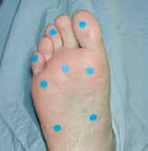
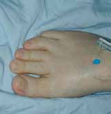
10g monofilament testing
The 10g monofilament testing is recommended as a screeningtool
to determine the presence of protective sensation in people with
· Loss of protective
· Reduced sweating
· Dysfunction of the
motor nerves that
· No perception of
Places for testing
shoes rubbing or
· Increased blood
foot. Limited joint
· Plantar surface of the metatarsal heads
increase plantar
(min. 3 metatarsal heads)12,13
· The great toe/first toe12
· Foot deformities
· The medial and lateral sides of the
plantar aspect of the midfoot13
· The plantar area of the heel13
· Unaware of a foot
· Dry skin with
· The dorsal aspect of the midfoot13
ulcer or lack of
longitudinal arch,
discomfort when a
· Bounding pulses
metatarsal heads
· Dilated dorsal
The pictures show testing sites
"There is no clear evidence on how many negative response sites equals an at-risk foot. Some literature shows that even one site with a negative response on each foot may indicate an at-risk foot." Baker, N. et al., 2005.12
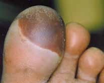
Areas at risk for neuropathic, ischaemic
Clinical symptoms of neuropathic
and neuro-ischaemic ulcers
and ischaemic foot ulcers14
In a cross-sectional, population-based study the proportion
of the lesions were*2
Clawed toes, possible high arch,
No specific deformities.
possible Charcot deformities
Possible absent toes/forefoot
from previous amputations
Warm, palpable pulse
Cold or decreased
temperature, pulse may
be absent or reduced
Pale/bluish. Pronounced redness
when lowered (dependent rubor),
blanching on elevation
Dry skin due to decreased
Thin, fragile and dry
Neuropathic ulcers 55% of total
Ischaemic ulcers 10% and
diabetic foot ulcers
neuro-ischaemic ulcers 34% of
On the plantar aspects (forefoot
Distal/tips of the toes,
total diabetic foot ulcers
80%) of the foot/toes
heel, or margins of the foot
Commonly seen on the
Not usual y. If present,
weight-bearing areas and is
distal eschar or necrosis
"Recent experience from our clinic indicates that the frequency of neuropathic ulcers has decreased, and the incidence of ischaemic
Usual y painless, with a
Painful, especial y with
and neuro-ischaemic ulcers has increased, equaling 50-50%."
"punched out" appearance
necrosis or slough
Mike Edmonds, 2005.
(granulation or deeper base)
surrounded by cal us
Reduced or absent sensation to
Sensation may be present but
touch, vibration, pain, and
decreased if there is associated
Usual y not present
Present and often bounding.
Absent or markedly reduced
Dilated, prominent veins
*1% of the ulcers were considered not to be diabetes-related.
Burning, stinging, shooting and
stabbing (non-stimulus dependent)
Deep infection or Charcot joint
Length, width, depth and location, preferably with clinical photograph
· Black (necrosis)
· Yel ow, red, pink
Be aware that some signs (fever, pain, increased white blood count/
ESR) may be absent. Evaluate the ulcer for signs of infection,
inflammation and oedema. For more information, please see page 20
Copious, moderate, mild, none
Cal us and scale, maceration, erythema, oedema
Wound undermining, deep tissue infection
Unhealthy wound edge
Superficial and deep infection symptoms10,15,16
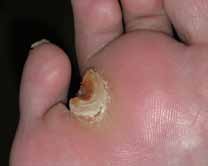
Wagner classification
Superficial (local) – Treat topically
No open lesions; may have deformity or cel ulitis
· Exuberant friable granulation tissue· Bright red discoloration of granulation tissue· Increased exudate· Malodour
Superficial diabetic ulcer (partial or ful thickness)
· New slough in wound base
Topical antimicrobial treatment may be considered for superficial/local infection, dependent on the assessment that wil direct the
Ulcer extension to ligament, tendon, joint capsule,
treatment. Superficial/local infection may, however, require
or deep fascia without abscess or osteomyelitis
systemic antibiotics. For further details and updates, please see the International Consensus on the Diabetic Foot, 2011.2
Deep – Treat systemically
Deep ulcer with abscess, osteomyelitis, or joint
· Pain· Probes to bone (increased risk in the
presence of osteomyelitis)
· New areas of break-down
Gangrene localised to portion of forefoot or heel
· Warmth· Erythema, oedema
Extensive gangrenous involvement of the entire
Signs of local and deep infection are potential y limb and/or life threatening. These clinical signs and symptoms require
Further reading:
urgent medical attention
International Consensus on the Diabetic Foot, The International Working Group on the Diabetic Foot, 20112, www.iwgdf.org
diabetic foot ulcerTreatment of diabetic foot ulcers
Local wound treatment
· If inadequate circulation, refer to vascular assessment and investigations
· Sharp surgery preferred
· Consider angioplasty, bypass or amputation
· Hydrogels, alginates and enzymes
Bacterial swabs help to identify organisms and sensitivity, but do not
diagnose infection in isolation from clinical features
Dependent on the outcomes of the wound assessment:
· Superficial/local – consider topical antimicrobial treatment (e.g. sustained
silver releasing dressings).
· Topical antimicrobials (e.g. sustained silver releasing dressings)
However, it may need systemic antibiotic therapy.
The general treatment may also include debridement of devitalized
· Systemic antibiotic therapy
tissue, pressure relief, optimising metabolic control and vascular
· Foams, alginates
· Deep – requires systemic antibiotic therapy to initial y cover
Gram-positive, Gram-negative and anaerobic organisms. Subsequently,
· The treatment of the edge depends on the outcomes of the
systemic antibiotic therapy can be modified according to the results of
assessment of the edge of the wound. In general, healthy
the culture. In addition, it is essential to consider the need for surgical
wounds have a pink woundbed and an advancing wound margin,
debridement, drainage of infection alongside pressure relief and
while un-healthy wounds have a dark and undermined wound
optimising metabolic control
· Topical antimicrobial (e.g. sustained silver-releasing dressings) may give
Occasional y, neuropathy can be associated with pain. For people
added benefit together with systemic coverage for deep infection
with painful diabetic neuropathy, consider the fol owing treatment:
· Appropriate offloading must be provided
Tricyclic antidepressants7,17 (TCAs):
· Total contact cast or pneumatic walker
· Second generation TCA agents17 e.g. duloxetine
· Deep toed or special shoes and orthotics
· First generation TCA agent7,17: amitriptyline
· Anticonvulsants: pregabalin17
Frequent (dependent on the clinical situation)
Application of moisture retentive dressings in the context of ischaemia and/or dry gangrene can result in a serious life-or-limb-
inspection of the diabetic foot ulcer is vital
threatening infection11
due to the increased risk of infection
Infection control is of paramount importance in DFU treatment because of its strong association with amputation. A study of 1,666 patients with diabetes found that foot infection increased the risk of amputation by 155 times19
Disclaimer: These are general guidelines. Please check local treatment recommendations applicable for your country or healthcare institution.
Disclaimer: These are general guidelines. Please check local treatment recommendations applicable for your country or healthcare institution.
Coloplast solutions for diabetic foot ulcers
Biatain® – superior absorption for faster wound healing
Biatain Soft-Hold – superior absorption for
Biatain is a soft and conformable foam dressing that effectively
wounds that are difficult to bandage
absorbs and retains wound exudate.20,21 This ensures a moisture
Biatain Soft-Hold has a gentle adherent layer
balance that is optimal for healing of exuding wounds.22,23
covering less than 50% of the foam surface al ows both hands to be free during dressing application and removal
Unique 3D polymer structure
SeaSorb® Soft – superior absorption for slough and cavity fillingHighly absorbent alginate dressing for moderately to heavily exuding wounds of any size and shape. Faster wound healing by conforming to any wound shape and by debridement of slough
Purilon® Gel – faster wound healing by effective
Biatain Non-Adhesive – superior absorption
and gentle debridement
for wounds with extra fragile skin
· Fast and effective debridement
Biatain Non-Adhesive is a soft and flexible absorbent
· High cohesion – the gel stays in place
polyurethane foam dressing with bevelled edges
Atrac-Tain® moisturizing cream
Biatain Silicone – superior absorption for
Atrac-Tain moisturizing cream is beneficial in the
treatment of moderate-to-severe xerosis of the feet
Biatain Silicone is a soft and flexible absorbent foam
in patients with diabetes24
dressing with a gentle silicone adhesive only on the border leaving the foam free to absorb exudate and heal the wound
Coloplast antimicrobial dressings for infected diabetic foot ulcers and ulcers at risk of infection
Biatain® Ag – superior absorption for infected wounds
Biatain Ag Non-Adhesive – superior absorption
Sustained release of silver during the entire wear time
for infected wounds with extra fragile skin
Biatain Ag is a soft and conformable silver foam
· Optimal healing environment26-27
dressing that is proven to help infected wounds heal
· Rapid kil ing of bacteria28
· Designed to prevent wound infection
Biatain Silicone Ag – superior absorption for infected woundsBiatain Silicone Ag is a soft and flexible absorbent silver foam dressing with a gentle silicone adhesive border
SeaSorb® Ag – superior absorption for slough and cavity filling on infected woundsHighly absorbent antimicrobial alginate dressing for moderately to heavily exuding infected wounds or wounds at risk of infection. Faster wound healing by conforming to any wound shape and by debridement of slough.
· Designed to fight cavity wound infection· Effect on a broad range of bacteria
Physiotulle® AgPhysiotul e Ag is a silver-containing, non-occlusive, hydrocol oid-based wound contact layer
Bakker, K. et al. The year of the diabetic foot, Diabetes Voice, March 2005, Vol. 50(1): 11-14.
17. CG96 Neuropathic pain - pharmacological management: ful guideline, NHS, National Institute for
Health and Clinical Excel ence, 27 May 2010 (http://guidance.nice.org.uk/CG96/Guidance/pdf/
International Working Group on the Diabetic Foot, International Consensus on the Diabetic Foot,
18. Sibbald, R.G. et al. Dermatological aspects of wound care, Chapter 30, In: Krasner, D.L. et al., A
Jude, E. et al. Assessment of the diabetic foot. Chronic Wound Care: Chapter 58, In: Krasner,
Clinical Sourcebook for Healthcare Professionals, Third Edition, HMP Communications Inc., 2001:
D.L. et al., A Clinical Sourcebook for Healthcare Professionals, Third Edition, HMP
Communications Inc. 2001: 589-597.
19. Lavery et al. Diabetes Care 2006;29(6):1288–93.
Armstrong, D.G. et al. Diabetic foot infections: stepwise medical and surgical management. International Wound Journal, 2004, Vol. 1(2): 123-132.
20. Andersen et al. A randomized, control ed study to compare the ef ectiveness of two foam
dressings in the management of lower leg ulcers. Ostomy/Wound Management 2002;(48)8:34-41.
Wil iams, R. et al. The size of the problem: Epidemiological and economic aspects of foot problems in diabetes. In: Boulton, A.J.M. et al., The Foot in Diabetes, John Wiley & Sons, Ltd.,
21. Thomas et al. www.dressings.org/TechnicalPublications/PDF/Coloplast-Dressings-
Spraul, M. Education – can it prevent diabetic foot ulcers and amputations? In: Boulton, A.J.M. et
22. White R and Cutting KF. Modern exudate management: a review of wound treatments.
al., The Foot in Diabetes, John Wiley & Sons, Ltd., 2000: 111-120.
Reddy, M. Wound healing: The next milennium. Diabetic Microvascular Complications Today, May/
23. Romanel i et al. Exudate management made easy. Wounds International 2010;1(2).
June 2005: 25-27.
24. Pham et al. A prospective, randomized, control ed double-blind study of a moisturizer for xerosis
Inlow, S. et al. Best practices for the prevention, diagnosis, and treatment of diabetic foot ulcers,
of the feet in patients with diabetes. OstomyWound Management 2002;48(5):30-36.
Ostomy/Wound Management 2000, Vol. 46(11): 55-68.
25. Buchholtz. An in-vitro comparison of antimicrobial activity and silver release from foam dressings.
Frykberg, R.G. et al. A summary of guidelines for managing the diabetic foot. Advances in Skin &
Wounds UK 2009.
Wound Care 2005, Vol. 18(4): 209-213.
26. Jørgensen et al. The silver-releasing foam dressing, Contreet Foam, promotes faster healing of
10. Edmonds, M. et al. A Practical Manual of Diabetic Foot Care, Blackwel Science, Oxford 2004.
critical y colonised venous leg ulcers: a randomised, control ed trial. International Wound Journal 2005;2(1):64-73.
11. Registered Nurses' Association of Ontario 2005. Assessment and management of foot ulcers for
people with diabetes. Toronto, Canada: Registered, Nurses' Association of Ontario.
27. Münter et al. Ef ect of a sustained silver-releasing dressing on ulcers with delayed healing: the
CONTOP study. Journal of Wound Care. 2006;15(5):199-206.
12. Baker, N. et al. A user's guide to foot screening. Part 1: Peripheral neuropathy, The Diabetic Foot
2005, Vol. 8(1): 28-37.
28. Ip et al. Antimicrobial activities of silver dressings: an in vitro comparison. Journal of Medical
13. Browne, A.C. et al. The diabetic neuropathic ulcer: An overview. Ostomy/Wound Management,
1999. Vol. 45 (No. 1A: Suppl).
14. Edmonds, M.E. et al. Managing the Diabetic Foot, Blackwel Science, Oxford 2005.
15. Sibbald, R.G. et al. Preparing the Wound Bed 2003: Focus on infection and inflammation,
Ostomy/Wound Management, November 2003, Vol. 49(1): 24-51.
16. Sibbald, R.G. et al. Cost–ef ective faster wound healing of critical y colonized wounds with a
sustained release silver foam dressing, based upon the symposium "Bacteria, sustained release of silver and improved healing", An official satel ite symposium of the WUWHS 2004. Published at www.worldwidewounds.com December 2005.
Biatain® – superior absorption
for faster healing
Superior absorption for
Superior absorption for
Superior absorption for
non-infected wounds*
Biatain Soft-Hold
Biatain Silicone Ag
Biatain Ibu Non-Adhesive
Biatain Silicone Lite
Biatain Ag Non-Adhesive
Biatain Ibu Soft-Hold
Sacral jun.
23x23
Biatain Non-Adhesive
Biatain Ag Adhesive
* Can be used for all types of exuding wounds.
Other Coloplast products
for diabetic foot ulcers
moisturizing cream
After 30 years in wound care,we at Coloplast believe that absorption is the key to better healing. Our Biatain® portfolio brings superior absorption to daily wound care needs, making Biatain the simple choice for faster healing.
Coloplast develops products and services that make life easier for people with very personal and private medical conditions. Working closely with the people who use our products, we create solutions that are sensitive to their
special needs. We cal this intimate healthcare. Our business includes ostomy care, urology and continence care
and wound and skin care. We operate global y and employ more than 7,000 people.
The Coloplast logo is a registered trademark of Coloplast A/S. [YYYY-MM.] Al rights reserved Coloplast A/S, 3050 Humlebæk, Denmark.
Source: http://www.coloplast.com.ar/Documents/Wound/CPWSC_Biatain_Diabetic-foot-ulcers_Quickguide_A4.pdf
Ausbildungsunterlagen für den Feuerwehr Medizinischen Dienst (Stand Nov.2007) Feuerwehr Medizinischer Dienst (FMD) im OÖLFV Der Feuerwehr Medizinische Dienst zusammengestellt von OBI Ing. Mag. Werner Kreisl (Sachbearbeiter FMD im Abschnitt Enns) Definition FMD: Zur Sicherstellung der
Healthy Living Suite 53, 5th Floor Morris Towers149 Wickham Terrace Brisbane QLD 4000Phone: 07 3839 1077ABN 255 059 949 60 Email: [email protected] Website: www.healthwiseclinic.com.au ISSUE 2 - 2008 Boosting your fertility • David McLeod B.Ac. ND • Zam Martin Obviously, an important component of